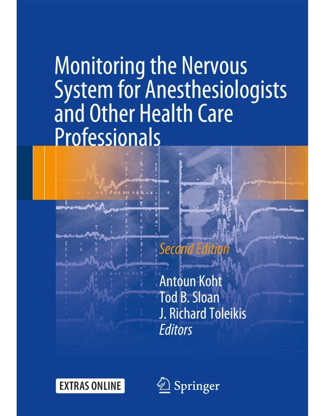
Monitoring the Nervous System for Anesthesiologists and Other Health Care Professionals
Livrare gratis la comenzi peste 500 RON. Pentru celelalte comenzi livrarea este 20 RON.
Disponibilitate: La comanda in aproximativ 4-6 saptamani
Editura: Springer
Limba: Engleza
Nr. pagini: 758
Coperta: Paperback
Dimensiuni: 17.78 x 4.06 x 25.4 cm
An aparitie: 26 Jun. 2017
Description:
This widely praised, first-of-its-kind book has been thoroughly updated, expanded, and enriched with extensive new case material, illustrations, and link-outs to multimedia, practice guidelines, and more. Written and edited by outstanding world experts, this was the first and remains the leading single-source volume on intraoperative neurophysiological monitoring (IOM). It is aimed at graduate students and trainees, as well as members of the operative team, including anesthesiologists, technologists, neurophysiologists, surgeons, and nurses.Now commonplace in procedures that place the nervous system at risk, such as orthopedics, neurosurgery, otologic surgery, vascular surgery, and others, effective IOM requires an unusually high degree of coordination among members of the operative team. The purpose of the book is to help students, trainees, and team members acquire a better understanding of one another’s roles and thereby to improve the quality of care and patient safety. From the reviews of the First Edition: “A welcome addition to reference works devoted to the expanding field of nervous system monitoring in the intraoperative period… will serve as a useful guide for many different health care professionals and particularly for anesthesiologists involved with this monitoring modality…An excellent reference…[and] a helpful guide both to the novice and to the developing expert in this field.” ‐‐Canadian Journal of Anesthesia “Impressive… [The book] is well written, indexed, and illustrated...The chapters are all extensively referenced. It is also very good value at the price....I would recommend this book to all residents and especially to all neuroanesthesiologists. It will make a worthwhile addition to their library.” ‐‐Journal of Neurosurgical Anesthesiology
Table of Contents:
Section I: Monitoring Techniques
1: Somatosensory-Evoked Potentials
Anatomy and Vascular Supply
Methods
Stimulation
Recording
Intraoperative Variables Affecting SSEPS: Pharmacology and Physiology
Inhalational Anesthetics
Intravenous Anesthetics
Temperature
Tissue Perfusion
Oxygenation/Ventilation
Intracranial Pressure
Other Physiologic Variables
Criteria for Intervention During Intraoperative SSEP Monitoring
Intraoperative Applications for SSEPs
Dermatomal-Evoked Potentials
References
2: Transcranial Motor-Evoked Potentials
Introduction
Motor Pathway Blood Supply
Technical Aspects of MEP Monitoring
Application of MEP Monitoring
Contribution of Anesthesiology to Effective MEP Monitoring
Risk of MEP Monitoring
Conclusion
References�
3: Auditory-Evoked Potentials
Anatomy of the Auditory System
Conduction of Auditory Signals from Ear to Cochlea
Neural Components of the Auditory System and Electrical Generators Along the Auditory Pathway
Cochlea: Electrocochleogram
Auditory Pathway from Cochlear Nerve to Midbrain: Auditory Brainstem-Evoked Responses
Primary Auditory Cortex: Mid-�Latency Auditory-Evoked Potentials
Vascular Supply of Auditory Pathway Structures
Techniques for Recording Auditory-Evoked Potentials
Stimulation
Electrocochleogram
Brainstem Auditory-Evoked Potentials
MLAEPs
Anesthetic and Physiologic Considerations for Monitoring of Auditory Brainstem Responses
References�
4: Visual-Evoked Potentials
Introduction
Anatomy and Physiology of the Visual System
Eliciting and Recording Flash Visual-Evoked Potentials
Flash Electrographic Response
Flash VEP Response
Techniques for Eliciting F-ERG and F-VEP
Devices for Flash Stimulation
Type of Light Stimulation
Monocular vs. Binocular Stimulation
Pupillary Size and Retinal Luminance
Recording F-ERGs and F-VEPs
Stimulus Color
Stimulus Rate
Recording Electrodes and Their Placement
Analysis Periods for F-ERGs and F-VEPs
Amplifier Settings for Recording F-ERG and F-VEP
Monitoring Criteria
Other IOM Applications with F-ERG and F-VEP
Retinal Stimulation and Intracranial Recording of Responses
Direct Electrical Stimulation of Optic Nerve
Effects of Temperature
Effects of Anesthesia on F-ERGs
Effects of Anesthesia on VEPs
Conclusion
References
5: Deep Brain Stimulation
Introduction
Surgery
The MER Procedure
Target Structures for the Case Examples
Cases and Disorders
Parkinson’s Disease
Procedure and Decisions
Dystonia
Procedure and Decisions
Status Dystonicus
Procedure and Decisions
Conclusion
References�
6: Monitoring of Spinal Cord Functions
Introduction
Recording Evoked SCPS for Intraoperative Monitoring
Recording Electric tc-MEPS and Magnetic tc-MEPS for Intraoperative Monitoring
Recording tc-SCPS for Intraoperative Monitoring
Anesthetics Used in Spinal Cord Monitoring
Case Studies (Clinical Applications)
Conclusion
Appendix: Techniques and Physiology
Introduction
The Catheter Electrodes
Origins of Each Component of the Segmental SCPs
References
7: Electromyography
Introduction
General Principle of EMG Monitoring
Cranial Nerve Monitoring Technique
Monitoring Specific Cranial Nerve Function
Cranial Nerves V and VII
Cranial Nerves III, IV, and VI
Cranial Nerves IX, X, XI, and XII
Brainstem Mapping
Mapping Technique
Anesthetic Management
Motor Strip Mapping
Techniques for Assessing Nerve Root Function and Pedicle Screw Placement
Methodology
Anesthetic Management
H-Reflex Testing
H-Reflex Response Acquisition
Anesthetic Management
References
8: The Use of Reflex Responses for IOM
Introduction
Anatomy and Neurophysiology
Spinal Cord Pathophysiology
Spinal Nerve Root Pathophysiology
Late Responses
H-Reflexes: Monosynaptic, Oligosynaptic
Neurophysiologic Basis of H-Reflexes
Gastrocnemius H-Reflex
Gastrocnemius H-Reflex Normal Parameters
Gastrocnemius H-Reflex Stimulation and Recording Techniques
Flexor Carpi Radialis H-Reflex
Flexor Carpi Radialis H-Reflex Background and Normal Parameters
Flexor Carpi Radialis H-Reflex Stimulation and Recording Techniques
Anesthetic Technique
Clinical Correlation of H-Reflexes and F-Responses
Clinical Correlation Summary
Polysynaptic Reflexes
Sacral Reflex
Intraoperative Application of BCRs
Monosynaptic and Polysynaptic Reflexes: Selective Dorsal Root Rhizotomy
Background
Technical Summary
Blink Reflex
Lower Extremity Intralimb and Interlimb Polysynaptic Reflexes
Conclusion
References
9: Brain and Spinal Cord Mapping
Introduction
Presurgical Mapping
Mapping During Awake Craniotomies
Location of the Central Sulcus by SSEP Polarity Reversal
Location of the Primary Motor Cortex with Electrical Stimulation
Subcortical Mapping
Spinal Cord Mapping
Conclusions
References�
10: EEG Monitoring
Introduction
The Genesis of the EEG
Cytoarchitecture
Control of Rhythm
EEG Signal Acquisition
Amplifiers and Filters
Signal Processing
Artifact Mitigation by Software
Time-Domain Methods
Clinical Applications of Time-Domain Methods
Burst Suppression and Its Quantitation
Frequency-Domain Methods
Spectral Displays in Clinical Practice
Bispectrum
Anesthetic Drugs Impact EEG
Volatile Agents
Nitrous Oxide
Barbiturates and Propofol
Etomidate
Ketamine
Narcotics
Benzodiazepines
Muscle Relaxants
References
11: Clinical Application of Raw and Processed EEG
Introduction
EEG Data Acquisition and Interpretation
Raw EEG
Processed EEG
Clinical Utility of Raw and Processed EEG
Future Directions of EEG Monitoring
Case Presentation
Conclusion
References
12: A Guide to Central Nervous System Near-Infrared Spectroscopic Monitoring
Introduction
Instrumentation
Transcranial NIRS Technology
Regional Cerebral Oxygen Saturation Measurement
Safety Considerations
Technical Considerations
Limitations of Cerebral Oximetry
Rationale for Cerebral NIRS Monitoring
Preoperative Factors
Systemic Arterial Pressure
Systemic Arterial Oxygenation
Systemic Arterial CO2 and pH
Cerebral Blood Flow Obstruction
Temperature
Anesthetic Adequacy
Seizure Activity
Supplemental Cerebral Perfusion
Rationale for Peri-spinal NIRS Monitoring
Conclusions
References�
13: Transcranial Doppler Ultrasound
Introduction
Technology
Principles of TCD Measurement
TCD Limitations
Rationale for Cerebral Hemodynamic Monitoring
Cerebral Blood Flow-Velocity Change
Cerebral Autoregulation
Vasomotor Reactivity
Neurovascular Coupling
Cerebral Embolization
Conclusion
References�
14: Monitoring of Jugular Venous Oxygen Saturation
Introduction
Rationale for SjvO2 Monitoring
Anatomy of Cerebral Venous Drainage
Technical Aspects of Jugular Venous Oximetry
Placement of Jugular Bulb Catheter
Continuous Fiberoptic Jugular Oximetry Versus Intermittent Sampling
Sampling Rate
Complications
Contraindications
Normative SjvO2 Values and Differential Diagnosis of Abnormal SjvO2 Values
Clinical Applications of SjvO2 Monitoring
Intensive Care Unit Uses of Jugular Venous Oximetry
Traumatic Brain Injury
Aneurysmal Subarachnoid Hemorrhage
Intracranial Arteriovenous Malformation
Intraoperative Uses of Jugular Venous Oximetry
Neurosurgical Anesthesia
Cardiac Surgery
Limitations
Conclusion
References�
Suggested Reading
15: Intracranial Pressure Monitoring
Introduction
References�
16: IOM Instrumentation Layout and Electrical Interference
Introduction
Basic Electronics and Definitions
Pathway 1: The Skin of the Patient Cskin
Pathway 2: Ambient Sources of Electrical Noise Vnoise
The Basic IOM Recording Circuit
Tissue Physiologic Generator (Vtissue)
Circuit Component: Tissue Between Generator and Electrode (Ztissue)
Circuit Component: Electrode Connection to the Patient + Electrode Components (Zelectrode)
Circuit Component: The Wire Between the Electrode Patient End and Connector End (Zleadwire)
Circuit Component: Amplifier Impedance (Za)
Circuit Component: Amplifier Iso-ground
Understanding Sources of Electrical Interference
Practical Tips
Specifically for the Anesthesiologist
For the IOM Professional
During Patient Setup
When Troubleshooting Electrical Noise
Technical Note: Modern IOM Equipment and Grounding
Recording Pathway: Input Switching
Recording Pathway: Amplifiers
Recording Pathway: Antialiasing Filters
Recording Pathway: A/D Convertors
Recording Pathway: Digital Signal and Computer Processing
Conclusion
References
17: Signal Optimization in Intraoperative Neuromonitoring
Patient-Related Issues
Somatosensory-Evoked Potentials
Peripheral Nerve Disorders
Central Nervous System Dysfunction
Motor-Evoked Potentials
Peripheral Nervous System Disorders
Central Nervous System Dysfunction
Electromyography and Nerve Conduction Studies
Electroencephalography
Anesthetic and Systemic Effects
Somatosensory-Evoked Potentials
Motor-Evoked Potentials
Electromyography
Electroencephalography
Technical Issues
Increased Electrical Noise
Electrical Noise: 60 Hz
Electrical Noise: High Frequency
Electrical Noise: Low Frequency or Intermittent
Electrical Noise: General Strategies
Poor Signal Amplitudes
Recording Technique
Stimulation Technique
Errors of Signal Acquisition
Electrode Plug-In Errors
System Errors
General Principles
Timing of Optimization
Prioritization
Conclusion
References�
Section II: Anesthesia Considerations
18: Anesthesia for Awake Neurosurgery
Introduction
Anesthesia and Monitoring for Awake Craniotomy During Tumor Resection
Indications and Patient Selection
Preoperative Evaluation and Preparation
Monitoring
Sedation and Analgesia
Regional Anesthesia
Anesthetic Management
Cortical and Subcortical Mapping and Neurologic Monitoring
Complications
Anesthesia for Awake Neurovascular Procedures
Anesthesia for Awake Deep Brain Stimulation
Anesthesia for Awake Seizure Surgery
Anesthesia for Awake Carotid Surgery
Sedation
Anesthesia
Neurologic Monitoring
Conclusion
References�
19: Anesthesia Management and Intraoperative Electrophysiological Monitoring
Impact of IOM Technique and Patient Comorbidity
Mechanism of Drug Action
Anesthetic Effects on the Electroencephalogram
General Effects
Specific Drugs and the EEG
Halogenated Agents
Nitrous Oxide
Propofol
Etomidate
Barbiturates
Benzodiazepines
Dexmedetomidine
Droperidol
Opioids
Ketamine
Anesthesia Selection and EEG
Anesthetic Effects on Evoked Potentials
Effect Based on Location of Synapses
Effect Based on Anesthesia Goals
Amnesia
Unconsciousness
Immobility
Antinociception
Specific Anesthesia Drugs
Halogenated Inhalational Agents
Nitrous Oxide
Intravenous Agents
Sedative-Hypnotics: Propofol
Etomidate
Benzodiazepines
Dexmedetomidine
Barbiturates
Droperidol
Intravenous Antinociception: Opioids
Ketamine
Lidocaine, Magnesium, and Regional Anesthesia
Neuromuscular Blocking Agents
Motor-Evoked Potentials
Facial Nerve
Recurrent Laryngeal Nerve
Peripheral Nerve Monitoring and Pedicle Screw Testing
If NMBA Is Used
Total Intravenous Anesthesia
Physiological Considerations in Anesthesia Management
Conclusion
References�
Section III: Clinical Applications
20: Monitoring Applications and Evaluating Changes
Reference
21: Intraoperative Neurophysiological Monitoring for Intracranial Aneurysm Surgery
Introduction
Case Presentation 1: MCA Aneurysm
Which Monitors Should You Consider for the Aneurysm Clipping?
What Do We Expect the Selected Neuromonitoring Modalities to Monitor During This Surgery?
What are Some Drawbacks of Each of the Above Monitors in Relation to This Surgery?
What is the Interpretation of These Neurophysiologic Monitoring Waveforms?
What is the Cause of This Change?
What is the Cause of These Changes?
Case Presentation 2: ICA/Ophthalmic Aneurysm
Should Only Upper Extremity SSEPs and TCMEPs, Only Lower Extremity SSEPs and TCMEPs, or Both Uppe
Case Presentation 3: Basilar Apex Aneurysm
How Does This Aneurysm Location Influence the Choice of Neurophysiologic Monitoring Modalities to
What are the Most Likely Etiologies for This Signal Change?
References
22: Intracranial Arteriovenous Malformation Surgery
Introduction
Case Presentation
References
23: Intraoperative Neurophysiologic Monitoring During Surgery for Supratentorial Mass Lesions
Case: Resection of an Insular Glioma
Risks of Surgery for Insular Tumors and Other Supratentorial Mass Lesions
Preservation of Nonmotor Function Using Mapping and Monitoring Techniques
Motor Mapping and Monitoring
Monitoring Results and Surgical Intervention
Possible Causes of the MEP Change and the Role of the Surgical Interventions
Why Is Neurophysiologic Monitoring Useful?
Conclusion
References
24: Surgery for Infratentorial Mass
Introduction
Resection of a Meningioma in the Right Cerebellopontine Angle
Potential Problems and Structures at Risk
Monitoring
Neurophysiological Monitoring
References
25: Trigeminal Microvascular Decompression
Introduction
Presentation of Clinical Scenarios
Case #1
What was the Cause of This Change? Was it Surgical, Pharmacologic, Physiologic, Positional, or
Case #2
What Could Be the Cause?
Case #3
Is This a Significant Change?
What is the Cause of This Change?
Case #4
What Happened? Is This Related to the Remifentanil Infusion, a Cardiac Incident, Brainstem Manip
Case #5
What is the Problem and What Should Be Done?
Case #6
Conclusion
References
26: Surgery for Hemifacial Spasm
Introduction
Electrophysiology and Pathophysiology of HFS
Anatomy of the Facial Nerve and Etiology of HFS
Imaging in HFS
Operative Technique of Microvascular Decompression of the Facial Nerve
Anesthetic Considerations During MVD for HFS
Principles of Intraoperative Neuromonitoring for HFS
Monitoring for Complications
Case Illustrations
Case 1: Understanding the AMR During MVD of the Facial Nerve
Case 2: “Frozen Shoulder” or Adhesive Capsulitis of the Glenohumeral Joint after MVD in the
Case 3: Inability to Access Brainstem Due to Intravenous Fluid Overload
Case 4: Technical Difficulties with the Inserted Earpiece for Acquiring ABRs
Case 5: Hearing Loss as a Result of MVD for HFS
Case 6: Anesthesia and the AMR
Case 7: Stroke During MVD for HFS
Case 8: Facial Weakness and MVD for HFS
Case 9: Vestibular Nerve Dysfunction (After MVD for HFS)
Case 10: Dysphagia/Hoarseness Following MVD for HFS
References
27: Skull Base Surgery
Introduction
Perioperative Considerations
Goals of IOM During Skull Base Surgery
Modes of Neuromonitoring
Electromyography
Evoked Potentials (SSEP, MEP,VEP, ABR)
Case 1
Differential Diagnosis of ABR Findings
Case Progression
Case 2
Differential Diagnosis of SSEP Findings
Case Progression
Conclusion
References
28: Surgery for Chiari Type I Malformation
Introduction
Anatomy and Pathophysiology
Symptomatology of Chiari I Malformation
Diagnosis of Chiari I Malformation
Operative Technique for Chiari I Malformation
Potential Structures at Risk During Surgery for Chiari I Malformation
Positioning Considerations During Surgery
Principles of Intraoperative Neuromonitoring for Chiari I Malformation Surgery
Case Illustration
Stroke During an Operation for Chiari I Malformation
Team Notes
References
Suggested Reading
29: ENT and Anterior Neck Surgery
Introduction
Outcomes
References
30: Carotid Surgery
Introduction
Case 1
Preoperative: Anesthetic and Neuromonitoring Considerations
Specific Goals in the Anesthetic Management of a Carotid Endarterectomy
Different Possible Monitoring Techniques to Assess Cerebral Blood Flow
Intraoperative Course
Transcranial Doppler Ultrasonography
Case 2
EEG Monitoring
Analysis of the Cause of Intraoperative Neuromonitoring Changes
Management of Changes
Outcome
Conclusion
References
31: Anterior Cervical Spine Surgery
Introduction
Case 1
Case 2
Case 3
Conclusion
References�
32: Posterior Cervical Spine Surgery
Introduction
Case Presentation
Discussion
References�
33: Surgery for Scoliosis Correction
Introduction
Case Report
Anesthetic
Discussion
Discussion
Discussion
Discussion
Discussion
Discussion
Discussion
Discussion
Discussion
Discussion
Discussion
References
34: Neurophysiological Monitoring in Thoracic Spine Surgery
Monitoring techniques for the prevention of blindness in the prone position are not �currently eff
Surgical Approach
Neurological Risks of Thoracic Surgery
Monitoring in Thoracic Spinal Surgery
Case 1
Discussion
Case 2
Discussion
Conclusions
References�
35: Intraoperative Neurophysiologic Monitoring for Lumbosacral Spine Procedures
Decompressive Laminectomy With or Without Fusion and Instrumentation
Lumbar Interbody Fusion
Posterior Lumbar Interbody Fusion (PLIF)
Transforaminal Lumbar Interbody Fusion (TLIF)
Anterior Lumbar Interbody Fusion (ALIF)
Extreme Lateral Interbody Fusion (ELIF/XLIF)
Paracoccygeal Transsacral Fixation
Lumbar Microdiscectomy
References�
36: Intramedullary Spinal Cord Surgery
Introduction
Neurophysiology
Anesthesia for Neurophysiologic Monitoring
Case 1
History, Clinical Assessment, and Imaging
Surgery and Intraoperative Monitoring
Case Summary Interpretation and Discussion
Case 2
History, Clinical Assessment, and Imaging
Surgery and Intraoperative Monitoring
Case Summary Interpretation and Discussion
Case 3
History, Clinical Assessment, and Imaging
Surgery and Intraoperative Monitoring
Summary, Interpretation, and Discussion
Discussion
References
Suggested Reading
37: Intraoperative Monitoring in Tethered Cord Surgery
Introduction
Case Presentation
Monitoring Modalities
Anesthetic Management
Intraoperative Neuromonitoring
Somatosensory-Evoked Potentials
Transcranial Motor-Evoked Potentials
Electromyography
Sensory-Evoked Potentials
Bladder Pressure Urometry
Bulbocavernosus Reflex
Intraoperative Course
Postoperative Outcome
References�
38: Surgery in the Peripheral Nervous System
Objectives
Practical Illustrations of the Application of Operative Recordings
Case 1
Case 2
Summary
References�
39: Surgery of the Aortic Arch
Case Presentation
Introduction
Hypothermia and Cerebral Protection During Circulatory Arrest
Temperature Monitoring
Neuroelectrophysiological Monitoring
Acid Base Management on Bypass
Cerebral Oximetry
Transcranial Doppler
Combination of Cerebral Oximetry and Transcranial Doppler
Conduct of the Surgical Repair of the Aortic Dissection
Conclusion
References�
40: Electrophysiological Monitoring During Thoracic Aortic Aneurysm Surgery
Introduction
IOM, Time, and Neurological Injury
Spinal Cord Blood Supply
Open Surgical TAA Repair
Monitoring Strategy During Open TAA Surgery
Application of Intraoperative Monitoring to TAA Surgery
Comparison of SSEP and MEP in Aorta Surgery
Endovascular Stent Placement
Hybrid Techniques
IOM Techniques During Thoracic Aortic Aneurysm Surgery
Presentation of Case 1
Discussion
Presentation of Case 2
Discussion
References�
41: Monitoring During Cardiopulmonary Bypass
Introduction
Causes of Injury During CPB and Neural Structures at Risk
Case Description
Available Physiologic Monitors
Monitor Noteworthy Changes
Differential Diagnosis
Technical
Physiologic
Pharmacologic
Surgical
Patient Management and Outcome
Conclusion
References�
42: Interventional Neuroradiology
Introduction
Neuromonitoring Plan
Anesthetic Management
Interpretation of Data
Case Studies
Conclusions
References�
43: Intraoperative Neuromonitoring in Pediatric Surgery
Introduction
Anesthetic Management of Pediatric Surgical Procedures That Require Intraoperative Neurophysiologi
Monitoring Modalities and Development
Somatosensory Evoked Potentials
Transcranial Motor Evoked Potentials
Electromyography
Electroencephalography
Brain Stem Auditory Evoked Responses
Bulbocavernosus Reflex
Common Pediatric Surgical Procedures Utilizing IONM
Posterior Spinal Fusion
Anesthetic Management During Spine Surgery
Dorsal Rhizotomy
Tethered Cord Release
Craniotomy for Tumor/Mass Lesion Resection
Conclusion
References
Suggested Reading
Section IV: Intensive Care
44: Monitoring in the Intensive Care Unit
Introduction
Case 1: Traumatic Brain Injury
Cerebral Oxygenation
Temperature Monitoring
Case 2: Subarachnoid Hemorrhage
Blood Pressure Monitoring
Case 3: Spinal Cord Injury
Monitoring Fluid Status and Cardiac Output
Pulmonary Artery Catheters
Preload Responsiveness
Conclusion
References�
45: Epilepsy and Seizures: OR and ICU Applications of EEG
Introduction
Epilepsy
Intensive Care EEG Monitoring
Other EEG Applications in the ICU
Conclusion
References�
46: Monitoring Cerebral Blood Flow
Physiology
Mechanisms of Cerebral Blood Flow Regulation
Autoregulation
Cerebrovascular Reserve
Cerebral Blood Flow Measurement and Clinical Applications of Specific Techniques Perioperatively
Stable Xenon CTCBF
Xe133 CBF
Jugular Bulb AVO2 Difference
Thermodilution rCBF
Transcranial Doppler
Vasospasm
Cerebrovascular Reserve
Intracranial Pressure
Brain Death
Vessel Patency
Emboli
Hyperemia
Near-Infrared Spectroscopy Based Monitors of CBF
Arterial Spin Labeling
Computed Tomography Perfusion
Intraoperative Cerebral Blood Flow Monitoring
Indocyanine Green Videoangiography
Monitors of Autoregulation
TCD-Based Autoregulation (Mx)
ICP-Based Autoregulation (Prx)
COx
Conclusion
References
Afterword to First Edition
Future of Monitoring the Nervous System
References
Afterword to First Edition
Future of Monitoring the Nervous System
References
| An aparitie | 26 Jun. 2017 |
| Autor | Antoun Koht, Tod B. Sloan, J. Richard Toleikis |
| Dimensiuni | 17.78 x 4.06 x 25.4 cm |
| Editura | Springer |
| Format | Paperback |
| ISBN | 9783319465401 |
| Limba | Engleza |
| Nr pag | 758 |


Clientii ebookshop.ro nu au adaugat inca opinii pentru acest produs. Fii primul care adauga o parere, folosind formularul de mai jos.