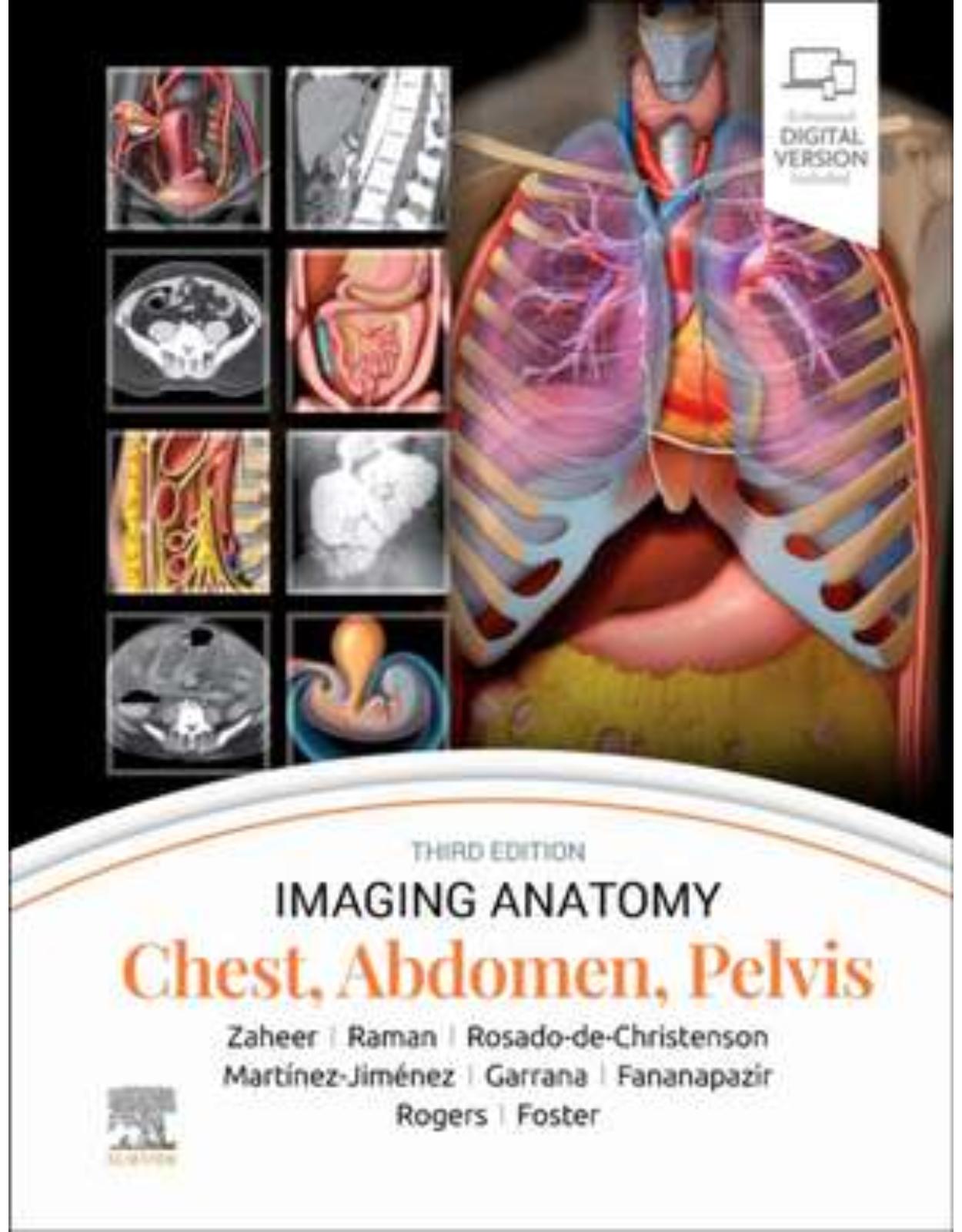
Imaging Anatomy: Chest, Abdomen, Pelvis
Livrare gratis la comenzi peste 500 RON. Pentru celelalte comenzi livrarea este 20 RON.
Disponibilitate: La comanda in aproximativ 4-6 saptamani
Editura: Elsevier
Limba: Engleza
Nr. pagini: 1192
Coperta: Hardcover
Dimensiuni: 215 x 276 mm
An aparitie: 16 oct 2023
|
Description: |
||
|
|
||
|
||
|
|
||
|
Section 1: Chest 4 Chest Overview Melissa L. Rosado de Christenson, MD, FACR 44 Lung Development Melissa L. Rosado de Christenson, MD, FACR 64 Airway Structure Brett W. Carter, MD, CPPS and Gerald F. Abbott, MD, FACR 86 Vascular Structure Melissa L. Rosado de Christenson, MD, FACR 106 Interstitial Network Brett W. Carter, MD, CPPS and Gerald F. Abbott, MD, FACR 118 Lungs Santiago Martínez-Jiménez, MD 150 Hila Melissa L. Rosado de Christenson, MD, FACR 180 Airways Brett W. Carter, MD, CPPS and Gerald F. Abbott, MD, FACR 202 Pulmonary Vessels Melissa L. Rosado de Christenson, MD, FACR 234 Pleura Brett W. Carter, MD, CPPS and Gerald F. Abbott, MD, FACR 260 Mediastinum Melissa L. Rosado de Christenson, MD, FACR 296 Systemic Vessels Melissa L. Rosado de Christenson, MD, FACR 338 Heart Melissa L. Rosado de Christenson, MD, FACR 382 Coronary Arteries and Cardiac Veins Akram M. Shaaban, MBBCh 404 Pericardium Melissa L. Rosado de Christenson, MD, FACR 424 Chest Wall Brett W. Carter, MD, CPPS and Gerald F. Abbott, MD, FACR Section 2: Abdomen 450 Embryology of Abdomen Siva P. Raman, MD 486 Abdominal Wall Siva P. Raman, MD 510 Diaphragm Siva P. Raman, MD 530 Peritoneal Cavity Siva P. Raman, MD 552 Vessels, Lymphatic System and Nerves, Abdominal Siva P. Raman, MD 594 Esophagus Siva P. Raman, MD 610 Gastroduodenal Siva P. Raman, MD 638 Small Intestine Atif Zaheer, MD and Siva P. Raman, MD 668 Colon Siva P. Raman, MD 710 Spleen Siva P. Raman, MD 734 Liver Siva P. Raman, MD 780 Biliary System Siva P. Raman, MD 806 Pancreas Siva P. Raman, MD 836 Retroperitoneum Siva P. Raman, MD 862 Adrenal Siva P. Raman, MD 884 Kidney Siva P. Raman, MD 922 Ureter and Bladder SECTION 3: PELVIS 948 Vessels, Lymphatic System and Nerves, Pelvic Paula J. Woodward, MD and Akram M. Shaaban, MBBCh Male 976 Male Pelvic Wall and Floor Paula J. Woodward, MD and Akram M. Shaaban, MBBCh 1002 Testes and Scrotum Paula J. Woodward, MD and Akram M. Shaaban, MBBCh 1020 Prostate and Seminal Vesicles Paula J. Woodward, MD and Akram M. Shaaban, MBBCh 1038 Penis and Urethra Paula J. Woodward, MD and Akram M. Shaaban, MBBCh Female 1052 Female Pelvic Floor Paula J. Woodward, MD, Rania Farouk El Sayed, MD, PhD, and Akram M. Shaaban, MBBCh 1080 Uterus Paula J. Woodward, MD and Akram M. Shaaban, MBBCh 1106 Ovaries Douglas Rogers, MD |
||
| An aparitie | 16 oct 2023 |
| Autor | Siva P. Raman, Melissa L. Rosado-de-Christenson, Atif Zaheer, Santiago Martínez-Jiménez, Ghaneh Fananapazir, Sherief H. Garrana, Douglas Rogers, Bryan R. Foster |
| Dimensiuni | 215 x 276 mm |
| Editura | Elsevier |
| Format | Hardcover |
| ISBN | 9780443118005 |
| Limba | Engleza |
| Nr pag | 1192 |
| Versiune digitala | DA |


Clientii ebookshop.ro nu au adaugat inca opinii pentru acest produs. Fii primul care adauga o parere, folosind formularul de mai jos.