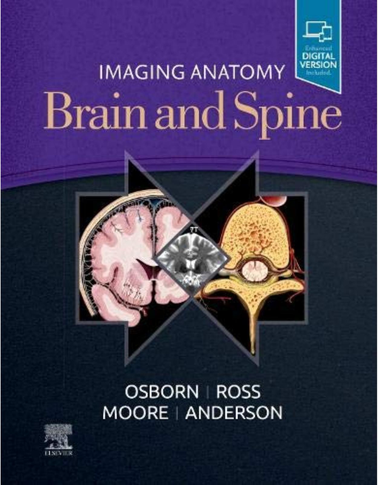
Imaging Anatomy Brain and Spine
Livrare gratis la comenzi peste 500 RON. Pentru celelalte comenzi livrarea este 20 RON.
Disponibilitate: La comanda in aproximativ 4-6 saptamani
Editura: Elsevier
Limba: Engleza
Nr. pagini: 928
Coperta: Hardcover
Dimensiuni: 23.5 x 4.4 x 29.2 cm
An aparitie: 22 July 2020
Description:
This richly illustrated and superbly organized text/atlas is an excellent point-of-care resource for practitioners at all levels of experience and training. Written by global leaders in the field, Imaging Anatomy: Brain and Spine provides a thorough understanding of the detailed normal anatomy that underlies contemporary imaging. This must-have reference employs a templated, highly formatted design; concise, bulleted text; and state-of- the-art images throughout that identify the clinical entities in each anatomic area.
Features more than 2,500 high-resolution images throughout, including 7T MR, fMRI, diffusion tensor MRI, and multidetector row CT images in many planes, combined with over 300 correlative full-color anatomic drawings that show human anatomy in the projections that radiologists use.
Covers only the brain and spine, presenting multiplanar normal imaging anatomy in all pertinent modalities for an unsurpassed, comprehensive point-of-care clinical reference.
Incorporates recent, stunning advances in imaging such as 7T and functional MR imaging, surface and segmented anatomy, single-photon emission computed tomography (SPECT) scans, dopamine transporter (DAT) scans, and 3D quantitative volumetric scans.
Places 7T MR images alongside 3T MR images to highlight the benefits of using 7T MR imaging as it becomes more widely available in the future.
Presents essential text in an easy-to-digest, bulleted format, enabling imaging specialists to find quick answers to anatomy questions encountered in daily practice.
Table of Contents
Copyright
Dedications
Contributing Authors
Preface
Acknowledgments
Sections:
Part I: Brain
SECTION 1: SCALP, SKULL, AND MENINGES
Chapter 1: Scalp and Calvarial Vault
Main Text
Image Gallery
Chapter 2: Cranial Meninges
Main Text
Image Gallery
Chapter 3: Pia and Perivascular Spaces
Main Text
Image Gallery
SECTION 2: SUPRATENTORIAL BRAIN ANATOMY
Chapter 4: Cerebral Hemispheres Overview
Main Text
Image Gallery
Chapter 5: Gyral/Sulcal Anatomy
Main Text
Image Gallery
Chapter 6: White Matter Tracts
Main Text
Image Gallery
Chapter 7: Basal Ganglia and Thalamus
Main Text
Image Gallery
Chapter 8: Other Deep Gray Nuclei
Main Text
Image Gallery
Chapter 9: Limbic System
Main Text
Image Gallery
Chapter 10: Sella, Pituitary, and Cavernous Sinus
Main Text
Image Gallery
Chapter 11: Pineal Region
Main Text
Image Gallery
Chapter 12: Primary Somatosensory Cortex (Areas 1, 2, 3)
Main Text
Image Gallery
Chapter 13: Primary Motor Cortex (Area 4)
Main Text
Image Gallery
Chapter 14: Superior Parietal Cortex (Areas 5, 7)
Main Text
Image Gallery
Chapter 15: Premotor Cortex and Supplementary Motor Area (Area 6)
Main Text
Image Gallery
Chapter 16: Superior Prefrontal Cortex (Area 8)
Main Text
Image Gallery
Chapter 17: Dorsolateral Prefrontal Cortex (Areas 9, 46)
Main Text
Image Gallery
Chapter 18: Frontal Pole (Area 10)
Main Text
Image Gallery
Chapter 19: Orbitofrontal Cortex (Area 11)
Main Text
Image Gallery
Chapter 20: Insula and Parainsula Areas (Areas 13, 43)
Main Text
Image Gallery
Chapter 21: Primary Visual and Visual Association Cortex (Areas 17, 18, 19)
Main Text
Image Gallery
Chapter 22: Temporal Cortex (Areas 20, 21, 22)
Main Text
Image Gallery
Chapter 23: Posterior Cingulate Cortex (Areas 23, 31)
Main Text
Image Gallery
Chapter 24: Anterior Cingulate Cortex (Areas 24, 32, 33)
Main Text
Image Gallery
Chapter 25: Subgenual Cingulate Cortex (Area 25)
Main Text
Image Gallery
Chapter 26: Retrosplenial Cingulate Cortex (Areas 29, 30)
Main Text
Image Gallery
Chapter 27: Parahippocampal Gyrus (Areas 28, 34, 35, 36)
Main Text
Image Gallery
Chapter 28: Fusiform Gyrus (Area 37)
Main Text
Image Gallery
Chapter 29: Temporal Pole (Area 38)
Main Text
Image Gallery
Chapter 30: Inferior Parietal Lobule (Areas 39, 40)
Main Text
Image Gallery
Chapter 31: Primary Auditory and Auditory Association Cortex (Areas 41, 42)
Main Text
Image Gallery
Chapter 32: Inferior Frontal Gyrus (Areas 44, 45, 47)
Main Text
Image Gallery
SECTION 3: BRAIN NETWORK ANATOMY
Chapter 33: Functional Network Overview
Main Text
Image Gallery
Chapter 34: Neurotransmitter Systems
Main Text
Image Gallery
Chapter 35: Default Mode Network
Main Text
Image Gallery
Chapter 36: Attention Control Network
Main Text
Image Gallery
Chapter 37: Sensorimotor Network
Main Text
Image Gallery
Chapter 38: Visual Network
Main Text
Image Gallery
Chapter 39: Limbic Network
Main Text
Image Gallery
Chapter 40: Language Network
Main Text
Image Gallery
Chapter 41: Memory Network
Main Text
Image Gallery
Chapter 42: Social Network
Main Text
Image Gallery
SECTION 4: INFRATENTORIAL BRAIN
Chapter 43: Brainstem and Cerebellum Overview
Main Text
Image Gallery
Chapter 44: Midbrain
Main Text
Image Gallery
Chapter 45: Pons
Main Text
Image Gallery
Chapter 46: Medulla
Main Text
Image Gallery
Chapter 47: Cerebellum
Main Text
Image Gallery
Chapter 48: Cerebellopontine Angle/IAC
Main Text
Image Gallery
SECTION 5: CSF SPACES
Chapter 49: Ventricles and Choroid Plexus
Main Text
Image Gallery
Chapter 50: Subarachnoid Spaces/Cisterns
Main Text
Image Gallery
SECTION 6: SKULL BASE AND CRANIAL NERVES
Chapter 51: Skull Base Overview
Main Text
Image Gallery
Chapter 52: Anterior Skull Base
Main Text
Image Gallery
Chapter 53: Central Skull Base
Main Text
Image Gallery
Chapter 54: Posterior Skull Base
Main Text
Image Gallery
Chapter 55: Cranial Nerves Overview
Main Text
Image Gallery
Chapter 56: Olfactory Nerve (CNI)
Main Text
Image Gallery
Chapter 57: Optic Nerve (CNII)
Main Text
Image Gallery
Chapter 58: Oculomotor Nerve (CNIII)
Main Text
Image Gallery
Chapter 59: Trochlear Nerve (CNIV)
Main Text
Image Gallery
Chapter 60: Trigeminal Nerve (CNV)
Main Text
Image Gallery
Chapter 61: Abducens Nerve (CNVI)
Main Text
Image Gallery
Chapter 62: Facial Nerve (CNVII)
Main Text
Image Gallery
Chapter 63: Vestibulocochlear Nerve (CNVIII)
Main Text
Image Gallery
Chapter 64: Glossopharyngeal Nerve (CNIX)
Main Text
Image Gallery
Chapter 65: Vagus Nerve (CNX)
Main Text
Image Gallery
Chapter 66: Accessory Nerve (CNXI)
Main Text
Image Gallery
Chapter 67: Hypoglossal Nerve (CNXII)
Main Text
Image Gallery
SECTION 7: EXTRACRANIAL ARTERIES
Chapter 68: Aortic Arch and Great Vessels
Main Text
Image Gallery
Chapter 69: Cervical Carotid Arteries
Main Text
Image Gallery
SECTION 8: INTRACRANIAL ARTERIES
Chapter 70: Intracranial Arteries Overview
Main Text
Image Gallery
Chapter 71: Intracranial Internal Carotid Artery
Main Text
Image Gallery
Chapter 72: Circle of Willis
Main Text
Image Gallery
Chapter 73: Anterior Cerebral Artery
Main Text
Image Gallery
Chapter 74: Middle Cerebral Artery
Main Text
Image Gallery
Chapter 75: Posterior Cerebral Artery
Main Text
Image Gallery
Chapter 76: Vertebrobasilar System
Main Text
Image Gallery
SECTION 9: VEINS AND VENOUS SINUSES
Chapter 77: Intracranial Venous System Overview
Main Text
Image Gallery
Chapter 78: Dural Sinuses
Main Text
Image Gallery
Chapter 79: Superficial Cerebral Veins
Main Text
Image Gallery
Chapter 80: Deep Cerebral Veins
Main Text
Image Gallery
Chapter 81: Posterior Fossa Veins
Main Text
Image Gallery
Chapter 82: Extracranial Veins
Main Text
Image Gallery
Part II: Spine
SECTION 1: VERTEBRAL COLUMN, DISCS, AND PARASPINAL MUSCLE
Chapter 83: Vertebral Column Overview
Main Text
Image Gallery
Chapter 84: Ossification
Main Text
Image Gallery
Chapter 85: Vertebral Body and Ligaments
Main Text
Image Gallery
Chapter 86: Intervertebral Disc and Facet Joints
Main Text
Image Gallery
Chapter 87: Paraspinal Muscles
Main Text
Image Gallery
Chapter 88: Craniocervical Junction
Main Text
Image Gallery
Chapter 89: Cervical Spine
Main Text
Image Gallery
Chapter 90: Thoracic Spine
Main Text
Image Gallery
Chapter 91: Lumbar Spine
Main Text
Image Gallery
Chapter 92: Sacrum and Coccyx
Main Text
Image Gallery
SECTION 2: CORD, MENINGES, AND SPACES
Chapter 93: Spinal Cord and Cauda Equina
Main Text
Image Gallery
Chapter 94: Meninges and Compartments
Main Text
Image Gallery
SECTION 3: VASCULAR
Chapter 95: Spinal Arterial Supply
Main Text
Image Gallery
Chapter 96: Spinal Veins and Venous Plexus
Main Text
Image Gallery
SECTION 4: PLEXI AND PERIPHERAL NERVES
Chapter 97: Brachial Plexus
Main Text
Image Gallery
Chapter 98: Lumbar Plexus
Main Text
Image Gallery
Chapter 99: Sacral Plexus and Sciatic Nerve
Main Text
Image Gallery
Chapter 100: Peripheral Nerve and Plexus Overview
Main Text
Image Gallery
| An aparitie | 22 July 2020 |
| Autor | Anne G. Osborn MD FACR, Karen L. Salzman MD, Jeffrey S. Anderson MD PhD , Arthur W. Toga, Meng Law MD, Jeffrey Ross MD , Kevin R. Moore MD |
| Dimensiuni | 23.5 x 4.4 x 29.2 cm |
| Editura | Elsevier |
| Format | Hardcover |
| ISBN | 9780323661140 |
| Limba | Engleza |
| Nr pag | 928 |
| Versiune digitala | DA |


Clientii ebookshop.ro nu au adaugat inca opinii pentru acest produs. Fii primul care adauga o parere, folosind formularul de mai jos.