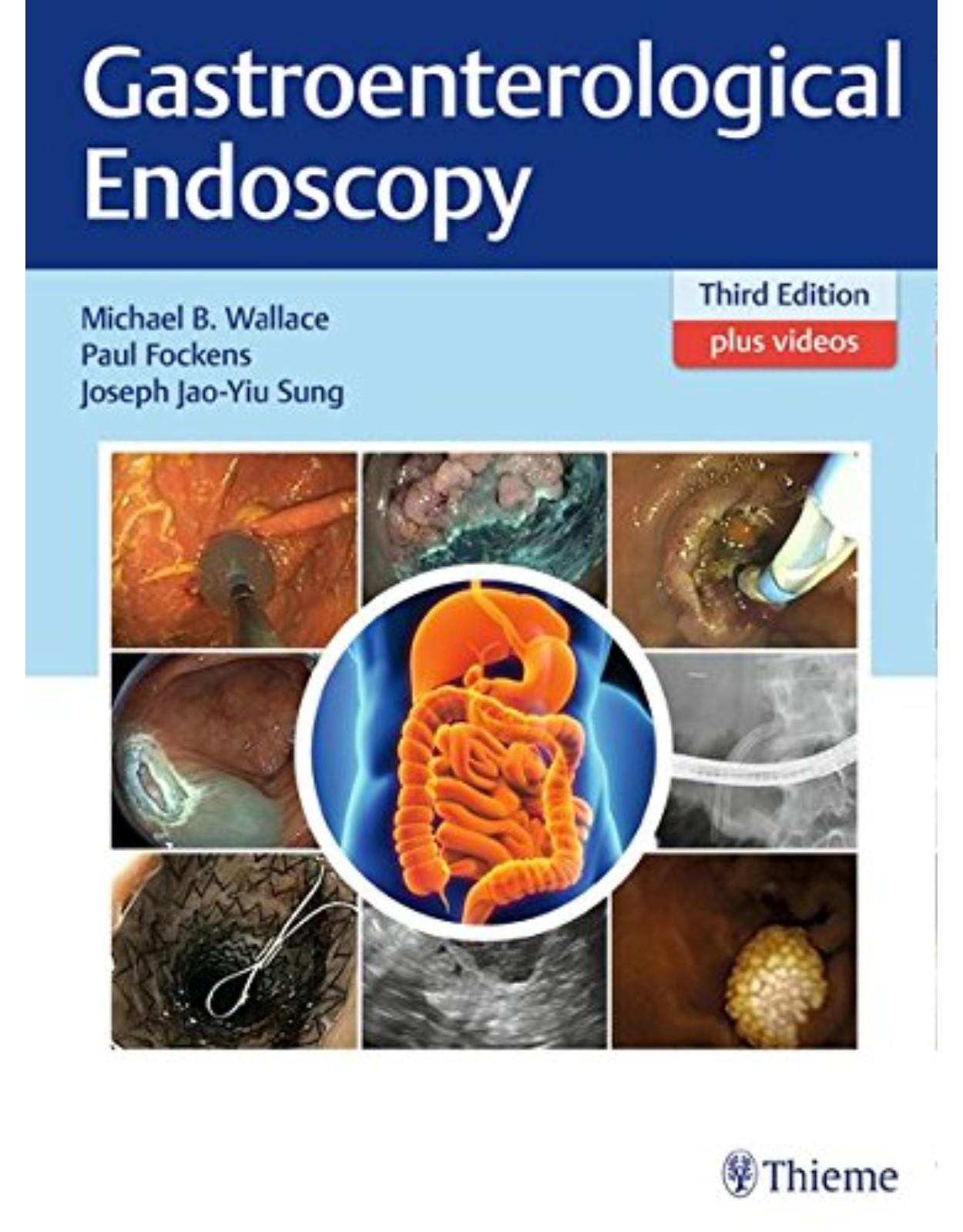
Gastroenterological Endoscopy
Livrare gratis la comenzi peste 500 RON. Pentru celelalte comenzi livrarea este 20 RON.
Disponibilitate: La comanda in aproximativ 4-6 saptamani
Editura: Thieme
Limba: Engleza
Nr. pagini: 438
Coperta: Hardback
Dimensiuni: 29.8 x 21.9 cm
An aparitie: 2018
Description:
Praise for the previous edition:
"Extraordinary achievement … this volume stands on its own as a marvelous feat in bringing such a vast array of clear and coherent instruction to endoscopists at all skill levels."—Gastroenterology
Written and edited by internationally renowned specialists, the third edition of Gastroenterological Endoscopy covers the entire spectrum of diagnostic and therapeutic procedures for the upper and lower GI tract while providing the latest overview of GI disorders. A great wealth of high-resolution photographs provides the visual information needed to confidently assess and diagnose mucosal lesions of the entire digestive tract. Significant advances in the field—both medical and technical—since the last edition are covered in comprehensive detail.
Key Features:
New panel of top international editors, continuing the tradition of excellence, depth, and breadth as originated by founding editors Classen, Tytgat, and Lightdale; list of contributing authors is a "who's who" of GI endoscopy
Coverage of newest, advanced tools and techniques: gastric-POEMS, submucosal tunnel endoscopic resection (STER), lumen-apposing metal stents, "over-the-scope" clips, and much more
More than 1,500 exquisite images
Gastroenterological Endoscopy, third edition, surely deserves a prominent place in any complete endoscopy reference collection.
Michael B. Wallace, MD, is Professor of Medicine at the Division of Gastroenterology and Hepatology, Mayo Clinic, Jacksonville, Florida, USA
Paul Fockens, MD, is Professor and Chair of the Dept. of Gastroenterology and Hepatology at the Academic Medical Center, University of Amsterdam, Amsterdam, The Netherlands
Joseph Jao-Yiu Sung, MD, PhD, is Mok Hing Yiu Professor of Medicine and Director of the Institute of Digestive Diseases of The Chinese University of Hong Kong, Shatin, Hong Kong
An award-winning international medical and scientific publisher, Thieme has demonstrated its commitment to the highest standard of quality in the state-of-the-art content and presentation of all its products. Founded in 1886, the Thieme name has become synonymous with high quality and excellence in online and print publishing.
Table of Contents:
I Introduction to Endoscopy
1 Education and Training in Endoscopy
1.1 Introduction
1.2 Clinical Education
1.2.1 Clinical Training to Competency in Esophagogastroduodenoscopy and Colonoscopy: Studies, Guidelines, and Assessment
1.2.2 Training in Endoscopic Retrograde Cholangiopancreatography
1.2.3 Complementary E-learning and Video Courses
1.3 Incorporation of Simulators in Training
1.4 Endoscopy Simulators and Training Models
1.4.1 Plastic Phantoms and Other Static Models
1.4.2. Computer Simulators
1.4.3 Training Courses with Live Animals
1.4.4 Ex Vivo Porcine Tissue Models (EASIE, Erlanger Endo-Trainer, EASIE-R)
1.4.5 Training Courses
1.4.6 Incorporating Simulator Training into Educational Programs and Maintaining Skills in Complex Procedures
References
2 The Value of Clinical Research
2.1 Introduction
2.2 Keys to Success
2.2.1 A Tough Skin
2.2.2 Building Teams
2.3 Designing Clinical Trials
2.3.1 Generating Ideas
2.3.2 Refining Ideas
2.3.3 Clinical Trial Design
2.3.4 Grant Writing
2.3.5 Conducting Clinical Trials
2.3.6 Presentation and National Meetings
2.3.7 Manuscript Writing
2.4 Ethics
2.4.1 Conflict of Interest
2.4.2 Registration of Clinical Trials and Underreporting of Negative Trials
2.4.3 Falsification of Data
2.4.4 Plagiarism
2.5 Manuscript Submission and Review Process
2.5.1 Expanding the Reach
2.5.2 The Future of Scientific Publications
References
II The Patient and Endoscopy
3 Informed Consent for Gastrointestinal Endoscopy
3.1 Introduction
3.2 What Is “Informed Consent”?
3.3 Clinician and Patient Relationship
3.4 What Information Is Required?
3.5 How Should the Information Be Provided?
3.6 Where and When Should the Consent Be Taken?
3.7 Withdrawal of Consent
3.8 Exceptions to the Requirement of Consent
References
4 Patient Preparation and Sedation for Endoscopy
4.1 Introduction
4.2 Presedation Assessment
4.3 Monitoring during Endoscopic Sedation
4.3.1 Introduction
4.3.2 Hemodynamic Monitoring
4.4 Pharmacology
4.4.1 Introduction
4.4.2 Benzodiazepines
4.4.3 Opioids
4.4.4 Propofol
4.4.5 Who Should Perform Endoscopic Sedation?
4.5 Postprocedure Care
4.5.1 Monitoring during Recovery
4.5.2 Discharge
References
5 Design of the Endoscopy Suite
5.1 Introduction
5.2 General Questions and Considerations
5.3 Guidelines for Planning an Endoscopy Suite
5.4 Pathways for Patients, Staff, and Material
5.5 Location of the Unit
5.6 Number of Rooms
5.7 X-Ray Requirements
5.8 The Endoscopic Examination Room
5.8.1 Size of the Rooms
5.8.2 Equipment
5.8.3 Monitor Systems and Anesthesia
5.8.4 Video Integration and PC-Based Documentation
5.8.5 Endoscopes and Endoscopic Equipment
5.9 Endoscopic Ultrasound and Laser Treatment Room, Radiography Room
5.10 Preparation and Recovery Room
5.11 Cleaning and Disinfection Area
5.12 Staffing
References
6 Cleaning and Disinfection in Endoscopy
6.1 Introduction
6.2 Principles of Disinfection
6.2.1 Definitions
6.2.2 Application to Gastrointestinal Endoscopes
6.2.3 Liquid Chemical Germicides and Automated Endoscope Reprocessors
6.3 Transmission of Infection by Gastrointestinal Endoscopy
6.3.1 Transmission by Endoscopes with Elevators
6.3.2 Failure or Breach in Reprocessing
6.3.3 Unusual Organisms
6.4 Design and Oversight of Reprocessing Facilities
References
7 Electrosurgical Principles for Endoscopy
7.1 Introduction
7.2 Electrosurgical Principles
7.2.1 Electrical and Tissue Variables
7.2.2 Monopolar versus Bipolar Circuit
7.3 Electrosurgical Units and Waveforms
7.4 Practical Applications
7.4.1 Snare Polypectomy
7.4.2 Hot Biopsy
7.4.3 Sphincterotomy
7.4.4 Hemostasis
7.4.5 Miscellaneous
7.5 Electrosurgical Hazards and Safety
7.5.1 Unintended Burn Injury
7.5.2 Implanted Electromagnetic Devices
7.5.3 Bowel Explosion
7.6 Conclusion
References
8 Antibiotic Prophylaxis in Endoscopy
8.1 Introduction
8.2 Bacteremia Related to Endoscopic Procedures
8.2.1 Procedures Associated with Low Risk of Bacteremia
8.2.2 Procedures Associated with High Risk of Bacteremia
8.3 Antibiotic Prophylaxis for the Prevention of Infective Endocarditis
8.3.1 Antibiotic Prophylaxis for the Prevention of Procedural-Related Infections (Other Than IE)
8.4 EUS-FNA
8.5 Percutaneous Endoscopic Gastrostomy/Jejunostomy
8.6 Cirrhosis with GI Bleeding
8.7 Synthetic Vascular Grafts and Other Nonvalvular Cardiovascular Devices
8.8 Orthopaedic Prostheses
8.9 Patients Receiving Peritoneal Dialysis
References
9 Quality Assurance in Endoscopy
9.1 The Importance of Quality
9.2 Performance Measures
9.3 Practicalities of Measurement
9.3.1 Clinical Importance
9.3.2 Standardization
9.3.3 Practicality
9.3.4 Governance Infrastructure
9.3.5 Negative Aspects
9.4 Quality Improvement
9.5 Summary
References
10 Endoscopic Complications
10.1 Introduction
10.2 General Considerations
10.2.1 Cardiopulmonary and Sedation-Related Events
10.2.2 Infection
10.3 Upper Gastrointestinal Endoscopy
10.3.1 Diagnostic Upper Gastrointestinal Endoscopy
10.3.2 Therapeutic Upper Gastrointestinal Endoscopy
10.3.3 Management of Upper Gastrointestinal Perforation
10.3.4 Management of Upper GI Bleeding
10.4 Small Bowel Endoscopy
10.5 Colonoscopy
10.5.1 Perforation
10.5.2 Management of Colonic Perforation
10.5.3 Bleeding
10.5.4 Unusual Complications
10.6 ERCP
10.6.1 Bleeding
10.6.2 Perforation
10.6.3 Infections
10.6.4 Post-ERCP Pancreatitis
10.7 Other Techniques
10.7.1 EUS-Guided Celiac Block/Neurolysis
10.7.2 EUS-Guided Drainage of Pancreatic Fluid Collections
10.7.3 Peroral Endoscopic Myotomy
10.8 Conclusion
10.9 Key Points
References
11 Anticoagulation and Endoscopy
11.1 Introduction
11.2 Antithrombotics
11.2.1 Antiplatelet Agents
11.2.2 Anticoagulants Agents
References
III General Diagnostic and Therapeutic Procedures and Techniques
12 Upper Gastrointestinal Endoscopy
12.1 History of Upper Gastrointestinal Endoscopy
12.2 General Diagnostic Techniques
12.2.1 Indications
12.2.2 Contraindications
12.3 Preparation of the Patient
12.4 Sedation
12.5 Use of Antifoaming Agents and Antispasmotics
12.6 Procedural Steps for Upper Gastrointestinal Endoscopy
12.6.1 Insertion and Observation
12.6.2 Esophagus
12.6.3 Esophagogastric Junction
12.6.4 Stomach and Duodenum
12.6.5 Transnasal Upper Endoscopy
12.7 Common Pathologies for Upper Gastrointestinal Endoscopy
12.7.1 I: Upper Gastrointestinal Cancers
12.7.2 II: Upper Gastrointestinal Hemorrhage
12.7.3 III: GERD and Barrett’s Esophagus
12.8 Screening for BE
12.9 Surveillance for BE
12.10 Barrett’s Esophagus–Related Dysplasia
12.11 Complications of Upper Gastrointestinal Endoscopy
References
13 Enteroscopy Techniques
13.1 Introduction
13.2 Overview of Enteroscopy Procedures
13.2.1 Anatomical Characteristics of the Small Intestine
13.2.2 Classification and Principles of Device-Assisted Enteroscopy
13.2.3 Balloon-Assisted Enteroscopy (Double-Balloon Endoscopy/Single-Balloon Endoscopy)
13.2.4 Spiral Endoscopy
13.3 General Diagnostic Techniques
13.4 General Therapeutic Techniques
13.4.1 Hemostasis
13.4.2 Balloon Dilation
13.4.3 Polypectomy/Endoscopic Mucosal Resection
13.4.4 Retrieval of Foreign Bodies
13.5 Accessory Devices and Techniques
13.6 Indications for the Use of Device-Assisted Enteroscopy
13.6.1 Indications for Diagnostic Use
13.6.2 Indications for Follow-Up of Small Intestinal Lesions
13.6.3 Therapeutic Indications for Device-Assisted Enteroscopy
13.6.4 Miscellaneous Indications for Device-Assisted Enteroscopy
13.7 Procedure-Specific Quality Measures
13.8 Procedure-Specific Training Requirements
13.8.1 Minimizing Air Insufflation for Deep Intubation
13.8.2 Necessity of X-Ray Fluoroscopy during Device-Assisted Enteroscopy
13.9 Minimizing Procedure-Specific Complications
13.9.1 Complications of Balloon-Assisted Endoscopy
13.9.2 Complications of Spiral Endoscopy
13.10 Conclusions
References
14 Wireless Video Capsule Endoscopy
14.1 Introduction
14.2 Technology
14.3 Setting and Preparation for Video Capsule Endoscopy
14.4 VCE Administration
14.5 Indications for VCE
14.6 Contraindications to VCE
14.7 Risk of VCE Retention
14.8 Reading a VCE Study
14.9 Conclusion
References
15 Colonoscopy: Preparation, Instrumentation, and Technique
15.1 Introduction
15.2 Preparation
15.2.1 Indications and Contraindications
15.2.2 Patient Preparation
15.3 Basic Instrumentation
15.3.1 Sedation
15.3.2 Colonoscope
15.3.3 Accessories
15.4 Technique
15.4.1 Scope Insertion
15.4.2 Scope Withdrawal
15.4.3 Polypectomy
15.4.4 Complications
15.5 Quality Measures
References
16 Endoscopic Retrograde Cholangiopancreatography
16.1 Introduction
16.2 Overview of Procedure
16.3 General Diagnostic Techniques
16.3.1 Biliary Cannulation
16.3.2 Sphincter of Oddi Manometry
16.4 General Therapeutic Techniques
16.4.1 Biliary Sphincterotomy
16.4.2 Endoscopic Papillary Balloon Dilation
16.4.3 Stone Extraction
16.4.4 Biliary Stenting
16.5 Accessory Devices and Techniques
16.5.1 Endoscopes
16.5.2 Equipment
16.6 Accepted Indications
16.7 Procedure-Specific Quality Measures
16.8 Procedure-Specific Training Requirements
16.9 Procedure-Specific Complications
References
17 Cholangioscopy
17.1 Introduction
17.2 Overview of Cholangioscopy
17.3 General Diagnostic Techniques
17.3.1 Two-Operator Systems: Mother-Baby Scopes
17.3.2 Single-Operator System: SpyGlass Cholangiopancreatoscopy
17.3.3 Direct Cholangioscopy
17.4 Accessory Devices and Techniques
17.4.1 Confocal Microscopy
17.4.2 Lithotripsy Probes
17.4.3 Intraductal Biopsy Forceps
17.5 Accepted Indications
17.5.1 Evaluation of Indeterminate and Malignant Biliary Strictures
17.5.2 Diagnosis and Management of Choledocholithiasis
17.5.3 Photodynamic Therapy of Cholangiocarcinoma
17.6 Complications of Cholangioscopy
References
18 Advanced Imaging Methods
18.1 Introduction
18.2 High-Definition Endoscopes
18.3 Virtual Chromoendoscopy
18.4 Narrow-Band Imaging
18.5 Flexible Spectral Imaging Color Enhancement
18.6 i-Scan and Optical Enhancement
18.7 Clinical Application of Virtual Chromoendoscopy
18.8 Chromoendoscopy
18.9 Clinical Application of Chromoendoscopy
18.10 Confocal Laser Endomicroscopy
18.11 Probe-Based CLE
18.12 Endoscope-Based CLE
18.13 Clinical Application
18.14 Optical Coherence Tomography
18.15 Conclusions
References
19 The Contribution of Histopathology to Endoscopy
19.1 Prerequisites
19.2 Clinical Impact of Histopathology by Segment within the Gastrointestinal Tract
19.2.1 Esophagus
19.2.2 Stomach
19.2.3 Small Bowel
19.2.4 Colorectum
19.3 Endoscopic Resections
19.4 Conclusion
References
20 Endoscopic Ultrasonography
20.1 Introduction
20.2 Overview of the Procedure
20.3 General Diagnostic and Therapeutic Techniques
20.3.1 Conditions of Implementation
20.3.2 Endoscopes and Probe
20.3.3 EUS Semiology of the Bowel Wall
20.3.4 Techniques
20.4 Accessory Devices and Techniques
20.4.1 Elastography
20.4.2 Contrast-Enhanced EUS
20.4.3 Needles and EUS-Guided Sample
20.5 Accepted Indications
20.6 Quality Measures
20.7 Training
20.8 Complications and Prevention
20.8.1 Noninterventional EUS
20.8.2 EUS-FNA
20.8.3 Interventional EUS
20.9 Prevention
References
21 Hybrid, Natural Orifice, and Laparoscopy-Assisted Endoscopy: New Paradigms in Minimally Invasive Therapy
21.1 Introduction
21.2 History of NOTES
21.3 Submucosal Surgery
21.4 Back to NOTES
21.5 Laparoscopy-Assisted Endoscopy
21.6 Laparoscopy-Assisted Endoscopic Resection
21.7 Endoscopy-Assisted Laparoscopic Resection
21.8 Combined Laparoscopic–Endoscopic Resection
21.9 Summary
References
IV Upper Gastrointestinal Tract Disease
22 Gastroesophageal Reflux Disease and Infectious Esophagitis
22.1 Diagnostic Approaches
22.2 Therapeutic Approaches
22.3 Surgical Therapy
22.4 Endoscopic Therapy for GERD
22.5 Infectious Esophagitis
22.5.1 Candida Esophagitis
22.5.2 Herpes Simplex Virus Esophagitis
22.5.3 Cytomegalovirus Esophagitis
22.5.4 Other Infections
References
23 Barrett’s Esophagus and Early Neoplasia
23.1 Diagnostic Work-Up for Barrett’s Esophagus and Early Neoplasia
23.1.1 General Approach to Barrett’s Esophagus
23.1.2 Endoscopic Imaging of Barrett’s Esophagus
23.2 Endoscopic Surveillance for Barrett’s Esophagus
23.3 Management of Dysplasia and Early Cancer in Barrett’s Esophagus
23.3.1 Indications for Endoscopic Treatment
23.3.2 Endoscopic Treatment Techniques
23.3.3 Current Guidelines for Endoscopic Treatment and Subsequent Follow-Up
23.4 Areas of Uncertainty, Experimental Techniques, and Research
23.4.1 Biological Markers in Barrett’s Esophagus
23.4.2 Low-Risk Submucosal Cancer
23.4.3 Novel Developments in Endoscopic Ablation
23.5 Conclusion
References
24 Squamous Neoplasia of the Esophagus
24.1 Introduction
24.1.1 Epidemiology and Risk Factors
24.1.2 Precursor Lesions for Squamous Neoplasia
24.2 Diagnostic Approaches
24.2.1 Nonendoscopic Techniques
24.2.2 Endoscopic Techniques
24.3 Treatment for Squamous Neoplasia of the Esophagus
24.3.1 Endoscopic Resection
24.4 Thermal Therapy
24.4.1 Radiofrequency Ablation
24.4.2 Cryotherapy
24.4.3 Other Ablative Techniques
24.5 Areas of Uncertainty, Experimental Techniques, and Research
24.6 Summary
References
25 Benign Esophageal Strictures and Esophageal Narrowing Including Eosinophilic Esophagitis
25.1 Introduction
25.2 Diagnostic Approaches
25.2.1 General Approach Including Causes, Symptoms, and Diagnosis
25.3 Classification System
25.4 Therapeutic Approaches
25.4.1 Standard Technique
25.4.2 Variations of Standard Techniques
25.5 Novel Diseases Causing Esophageal Stricturing
25.5.1 Eosinophilic Esophagitis
25.6 Post–Endoscopic Resection
25.7 Areas of Uncertainty, Experimental Techniques, and Research
References
26 Achalasia
26.1 Introduction
26.1.1 Epidemiology
26.1.2 Pathophysiology
26.1.3 Etiology
26.1.4 Clinical Presentation
26.2 Diagnostic Approaches
26.2.1 General Approach Including Equipment and Techniques
26.2.2 Achalasia Subtypes
26.2.3 Guidelines and Systematic Reviews
26.3 Therapeutic Approaches
26.3.1 Standard Techniques
26.3.2 Guidelines and Systematic Reviews
26.4 Areas of Uncertainty, Experimental Techniques, and Research
References
27 Advanced Esophageal Cancer
27.1 Introduction
27.2 Diagnosis and Classification
27.2.1 Malignant Dysphagia
27.2.2 Stents
27.2.3 Cryotherapy
27.2.4 Feeding Tubes
27.2.5 Esophagorespiratory Fistulas
27.2.6 Stents
27.2.7 Clips
27.2.8 Sutures
27.2.9 Bleeding
27.3 Conclusion
References
28 Peptic Ulcer Disease and Bleeding, Including Duodenal Ulcer
28.1 Introduction
28.2 Diagnosis of Peptic Ulcer Disease and Bleeding
28.3 Choice of Instrument for Peptic Ulcer Bleeding
28.4 Therapeutic Modalities for Peptic Ulcer Bleeding
28.4.1 Injection Therapy
28.4.2 Thermal
28.4.3 Mechanical Therapy
28.4.4 Topical Hemostatic Powders
28.5 New Hemostatic Modalities
28.5.1 Endoscopic Suturing
28.5.2 Endoscopic Ultrasound–Guided Angiotherapy
28.6 Conclusion
Reference
29 Gastric Cancer Including Early Neoplasia and Preneoplastic Conditions
29.1 Introduction
29.2 Diagnostic Approach
29.2.1 Preparation
29.2.2 Endoscopic Technique
29.2.3 Knowledge for Diagnosis
29.3 Therapeutic Approach
29.3.1 Principle of Endoscopic Resection
29.3.2 Indication for Endoscopic Resection
29.3.3 Clinical Management after Endoscopic Resection
29.4 Future Prospects
29.5 Repeat as Needed for Each Condition
References
30 Obesity: Endoscopic Approaches
30.1 Introduction
30.2 Obesity: Endoscopic Approaches
30.3 Diagnostic Approach and the Multidisciplinary Obesity Center Concept
30.3.1 General Approach, Equipment, and Techniques
30.3.2 Therapeutic Approaches: Currently Available Techniques
30.3.3 Gastric Techniques
30.3.4 Small Bowel Techniques
30.3.5 Endoscopic Revision of Prior Gastric Bypass
30.3.6 Other Postoperative Issues That Lead to Weight Gain and May Require Endoscopic Intervention
30.3.7 Guidelines and Systematic Reviews
30.3.8 Experimental Techniques
30.4 Summary
References
31 Small Intestinal Diseases Beyond the Duodenum
31.1 Introduction
31.2 Suspected Small Bowel Bleeding
31.2.1 Diagnostic Approaches
31.2.2 Therapeutic Approaches
31.3 Small Bowel Crohn’s Disease
31.3.1 Diagnosis
31.3.2 Therapeutics
31.4 Dilation of Small Bowel Stricture
31.5 Small Bowel Tumors
31.5.1 Diagnosis
31.5.2 Therapeutics
31.6 Malabsorption Disorders of the Small Bowel
31.6.1 Diagnostics and Therapeutics
31.7 Small Intestinal Infections
31.8 Congenital Lesions
31.9 Miscellaneous Conditions
31.10 Conclusion
References
32 Sporadic Neoplastic Polyps of the Duodenum and Ampulla
32.1 Introduction
32.2 Ampullary Neoplastic Polyps
32.2.1 Types of Ampullary Polyps
32.2.2 Clinical Manifestations
32.2.3 Diagnosis
32.2.4 Management of Ampullary Neoplasms
32.2.5 Endoscopic Ampullectomy
32.2.6 Endoscopic Outcomes: Clinical Success, Recurrence Rates
32.2.7 Surveillance
32.3 Conclusion
32.4 Nonampullary Sporadic Neoplastic Duodenal Polyps
32.4.1 Types of Nonampullary Duodenal Polyps
32.4.2 Diagnosis
32.4.3 Management of Nonampullary Duodenal Adenomas
32.4.4 Outcomes of Endoscopic Mucosal Resection
32.4.5 Adverse Events
32.4.6 Post–Endoscopic Mucosal Resection Care
32.4.7 Role of Endoscopic Submucosal Dissection
32.4.8 Surveillance
32.5 Conclusion
References
33 Malabsorption and Food Allergy/Intolerance
33.1 Introduction
33.2 Standard Endoscopy
33.2.1 Water-Immersion Technique
33.3 Chromoendoscopy and Magnification Endoscopy
33.4 Narrow-Band Imaging
33.5 Confocal Laser Endomicroscopy
33.6 Optical Coherence Tomography
33.7 Device-Assisted Enteroscopy
33.7.1 Capsule Endoscopy
33.8 Selected Small Bowel Diseases
33.8.1 Celiac Disease
33.8.2 Tropical Sprue
33.8.3 Small Bowel Bacterial Overgrowth
33.8.4 Sprue-Like Enteropathy Associated with Olmesartan
33.9 Conclusion
References
34 Portal Hypertension, Varices, Gastropathy, and Gastric Antral Vascular Ectasia
34.1 Introduction
34.2 Portal Hypertension: What Do We Need to Know?
34.2.1 Pathophysiology of Portal Hypertension
34.2.2 Noncirrhotic Portal Hypertension
34.2.3 Cirrhotic Portal Hypertension: Natural History, Risk Stratification, and Individualizing Care
34.3 Diagnosis of Portal Hypertension
34.3.1 Hepatic Venous Pressure Gradient
34.3.2 Noninvasive Tests
34.4 Treatment of Portal Hypertension
34.4.1 Primary Prophylaxis
34.4.2 Management of Acute Variceal Bleeding
34.5 Secondary Prophylaxis
34.6 Management of Treatment Failure
34.7 New Modality in Management of AVB: Hemospray
34.8 Management of Gastric Varices
34.9 Management of Ectopic Varices
34.10 Portal Hypertensive Gastropathy and Gastric Antral Vascular Ectasia
34.11 Areas of Uncertainty, Experimental Techniques, and Research
34.12 Conclusion
References
V Lower Gastrointestinal Tract Disease
35 Colorectal Polyps and Cancer Screening/Prevention
35.1 Introduction
35.2 Polyp Classification and Polyp Cancer Sequences
35.3 Conventional Adenomas
35.3.1 Low-Risk versus Advanced Conventional Adenomas
35.3.2 Shape and Colonic Distribution of Conventional Adenomas
35.3.3 Surface Features of Conventional Adenomas
35.3.4 Resection of Conventional Adenomas
35.4 Serrated Class Lesions
35.4.1 Terminology and Histology
35.4.2 Endoscopic Presentation
35.4.3 Resection of Serrated Lesions
35.5 Colorectal Cancer Screening
35.5.1 Approaches to Offering Screening
35.5.2 Factors That Affect Colorectal Cancer Risk
35.5.3 Choices of Individual Screening Tests
35.5.4 Surveillance after Cancer Resection
35.6 Conclusion
References
36 Advanced Colorectal Polyps and Early Cancer Resection
36.1 Introduction
36.2 Technical Aspects and Preparation
36.2.1 Patient Preparations
36.2.2 Techniques of Endoscopic Resection
36.2.3 Equipment Required
36.3 Lesion Assessment
36.4 Resection Technique
36.4.1 Endoscopic Mucosal Resection
36.4.2 Endoscopic Submucosal Dissection
36.5 Unique Situations
36.5.1 EMR of LSLs at the Anorectal Junction
36.5.2 EMR of LSLs at the Ileocecal Valve
36.5.3 EMR of Circumferential LSLs
36.5.4 EMR of Lumen Filling Lesions
36.5.5 EMR of Periappendiceal LSLs
36.5.6 EMR of Multiple Recurrent LSLs
36.5.7 Sessile Serrated Lesions
36.5.8 Endoscopic Resection of Large Pedunculated Lesions
36.6 Endoscopy versus Surgery
36.7 Complications
36.7.1 Intraprocedural Bleeding
36.7.2 Clinically Significant Postendoscopic Bleeding
36.7.3 Deep Injury
36.7.4 Postprocedural Pain
36.8 Residual and Recurrent Disease
36.8.1 Recurrence and EMR
36.8.2 Techniques at the Initial EMR to Prevent Recurrence
36.8.3 Triaging Patients to Follow Up Based on Risk of Recurrence
36.8.4 Accurate Assessment of the Post-EMR Scar
36.8.5 Endoscopic Treatment of Post-EMR Recurrence
36.9 Future Direction of ER
References
37 Inheritable Cancer Syndromes
37.1 Introduction
37.2 Nonpolyposis Syndromes
37.2.1 Lynch’s syndrome
37.2.2 Familial CRC
37.3 Polyposis Syndromes
37.3.1 Familial Adenomatous Polyposis
37.3.2 Attenuated Familial Adenomatous Polyposis
37.3.3 MUTYH-Associated polyposis
37.3.4 Serrated Polyposis Syndrome
37.3.5 Hamartomatous Polyposis Syndromes
References
38 Inflammatory Bowel Disease and Microscopic Colitis
38.1 Introduction
38.2 Endoscopic Characteristics of IBD
38.2.1 Lower Endoscopy
38.2.2 Upper Endoscopy
38.2.3 Small Bowel Imaging
38.2.4 Endoscopic Ultrasonography
38.2.5 Endoscopic Retrograde Cholangiopancreatography
38.3 Endoscopy in Established IBD
38.3.1 Acute Colitis
38.3.2 Routine Endoscopy
38.4 Endoscopic Evaluation of IBD Disease Activity
38.4.1 Crohn’s Disease
38.4.2 Ulcerative Colitis
38.5 Endoscopy after Surgery
38.5.1 Lower Endoscopy
38.6 Endoscopic Surveillance in IBD
38.7 Therapeutic Endoscopic Approaches in IBD
38.8 Microscopic Colitis
References
39 Lower Intestinal Bleeding Disorders
39.1 Introduction
39.2 General Aspects
39.2.1 Epidemiology
39.2.2 Clinical Course and Prognosis
39.3 Diagnostic Approach
39.3.1 History
39.3.2 Physical Examination
39.3.3 Laboratory Studies
39.3.4 Endoscopy
39.3.5 Nonendoscopic Methods
39.4 Differential Diagnosis
39.4.1 Diverticula
39.4.2 Vascular Diseases
39.4.3 Inflammation
39.4.4 Neoplasia
39.4.5 Anorectal Diseases
39.5 Therapy
39.5.1 Initial Resuscitation
39.5.2 Endoscopy
39.6 Injection Therapy
39.7 Thermocoagulation
39.8 Topical Agents
39.8.1 Hemospray (Cook Medical) TG 325
39.8.2 EndoClot (EndoClot Plus Inc.)
39.8.3 Ankaferd Blood Stopper (Ankaferd Health Products)
39.9 Mechanical Methods
39.9.1 Over-the-scope Clip System (OTSC) (Ovesco, Tübingen, Germany)
39.10 Differential Endoscopic Therapy
39.10.1 Diverticula
39.10.2 Vascular Diseases
39.10.3 Inflammation
39.10.4 Neoplasia
39.10.5 Bleeding after Colonic Polypectomy
References
40 Anorectal Diseases
40.1 Introduction
40.2 Inflammation
40.2.1 Crohn’s Disease
40.2.2 Perianal Abscesses
40.2.3 Anorectal Fistula
40.3 Infection
40.3.1 Chlamydial Infection
40.3.2 Gonococcal Proctitis
40.3.3 Herpes Simplex Virus
40.3.4 Syphilis
40.3.5 Lymphogranuloma Venereum
40.4 Vascular Cause
40.4.1 Ischemic Proctitis
40.4.2 Radiation Proctitis
40.5 Neoplasm
40.5.1 Anal Cancer
40.5.2 Anal Intraepithelial Neoplasia
40.6 Mechanical Cause
40.6.1 Hemorrhoids
40.6.2 Rectal Prolapse
40.6.3 Solitary Rectal Ulcer Syndrome
40.6.4 Anal Fissure
40.6.5 Stercoral Ulcer
References
VI Biliopancreatic, Hepatic, and Peritoneal Diseases
41 Benign Biliary Disorders
41.1 Introduction
41.2 Postoperative Biliary Stricture
41.3 Chronic Pancreatitis and Biliary Strictures
41.4 Primary Sclerosing Cholangitis
41.5 Bile Duct Leaks
References
42 Malignant Biliary Disease
42.1 Introduction
42.2 Diagnostic Approach
42.2.1 Radiologic Imaging
42.2.2 Endoscopic Retrograde Cholangiopancreatography
42.2.3 Fluorescence In-Situ Hybridization
42.2.4 Cholangioscopy
42.2.5 Endoscopic Ultrasound-Fine Needle Aspiration
42.2.6 Intraductal Ultrasound
42.2.7 Probe-based Confocal Laser Endomicroscopy
42.3 Classification Systems
42.4 Guidelines and Systematic Reviews
42.5 Therapeutic Approaches
42.5.1 Standard Techniques
42.5.2 Liver Transplantation
42.5.3 Variation of Standard Techniques
42.6 Guidelines and Reviews
42.7 Areas of Uncertainty
References
43 Acute and Chronic Pancreatitis
43.1 Introduction
43.2 Diagnostic Approaches
43.2.1 Overview
43.2.2 Equipment and Techniques
43.2.3 Guidelines and Systematic Reviews
43.3 Therapeutic Approaches
43.3.1 Standard Techniques
43.3.2 Guidelines and Systemic Reviews
43.4 Areas of Uncertainty, Experimental Techniques, and Research
43.4.1 Diagnostic Procedures
43.4.2 Therapeutic Procedures
43.5 Conclusion
References
44 Pancreatic Cancers and Cystic Neoplasms
44.1 Introduction
44.2 Pancreatic Cancers
44.2.1 Ductal Adenocarcinoma of the Pancreas
44.2.2 Pancreatic Neuroendocrine Tumors
44.3 Cystic Lesions of Pancreas
44.3.1 Nonneoplastic Cysts
44.3.2 Pancreatic Cystic Neoplasms
References
45 Subepithelial Tumors of the Gastrointestinal Tract
45.1 Introduction
45.2 Types of SETs
45.2.1 Gastrointestinal Stromal Tumors
45.2.2 Leiomyomas
45.2.3 Carcinoids
45.2.4 Other Subepithelial Lesions of the Gastrointestinal Tract
45.3 Risk Stratification of Subepithelial Tumors
45.4 Methods for Tissue Acquisition
45.4.1 Endoscopic Ultrasound-Guided Fine-Needle Aspiration
45.4.2 Endoscopic Ultrasound-Guided Fine-Needle Biopsy and Trucut Biopsy
45.4.3 Other Tissue Acquisition Techniques
45.5 Management of Subepithelial Lesions
45.6 Endoscopic Resection of Subepithelial Tumors
References
46 Gastrointestinal Foreign Bodies
46.1 Introduction
46.2 Clinical Epidemiology
46.2.1 Overview of Pathophysiology
46.3 Patient Presentation
46.4 Diagnosis
46.4.1 Treatment
46.4.2 Pharmacologic therapies
46.4.3 Endoscopic accessories and interventions
46.4.4 Esophageal food impaction
46.4.5 Sharp foreign bodies
46.4.6 Coins and button batteries
46.4.7 Bezoars
46.4.8 Rectal foreign bodies
46.5 Complications
46.6 Conclusion and Future Trends
References
Index
| An aparitie | 2018 |
| Autor | Michael B. Wallace, Paul Fockens, Joseph Jao-Yiu Sung |
| Dimensiuni | 29.8 x 21.9 cm |
| Editura | Thieme |
| Format | Hardback |
| ISBN | 9783131258533 |
| Limba | Engleza |
| Nr pag | 438 |


Clientii ebookshop.ro nu au adaugat inca opinii pentru acest produs. Fii primul care adauga o parere, folosind formularul de mai jos.