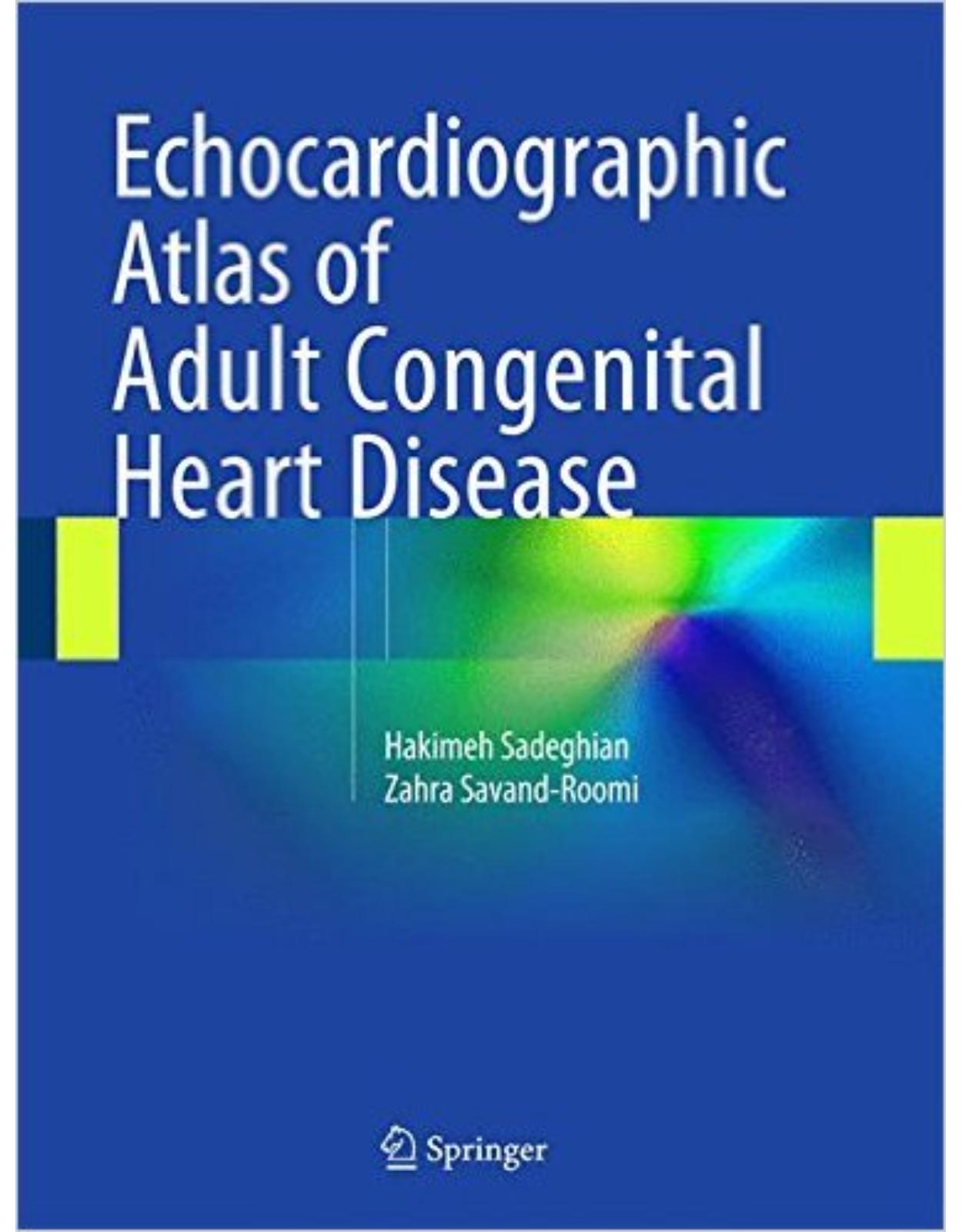
Echocardiographic Atlas of Adult Congenital Heart Disease 2015th Edition
Livrare gratis la comenzi peste 500 RON. Pentru celelalte comenzi livrarea este 20 RON.
Disponibilitate: La comanda in aproximativ 4 saptamani
Editura: Springer
Limba: Engleza
Nr. pagini: 517
Coperta: Hardback
Dimensiuni: 11.2 x 8.6 x 1.1 inches
An aparitie: 2015 edition (August 11, 2015)
Description:
This atlas of echocardiography presents more than 100 cases of adult congenital heart disease, from diagnosis to treatment follow-up. The coverage is broad, encompassing atrial and ventricular septal defects, patent ductus arteriosus, cyanotic adult congenital heart disease, and numerous other anomalies, as well as findings on fetal echocardiography. For each disease, all echocardiographic images and views which proved of diagnostic value are arranged sequentially, with inclusion of transesophageal echocardiographic images whenever appropriate. Additional pertinent information is provided relating to diagnosis and treatment, and key teaching points are highlighted. The superb quality of the illustrations and the range of cases considered (including many rare ones) ensure that this atlas will be of great value for cardiology residents and fellows and highly relevant to day-to-day practice.
Table of Contents:
Case 1: Atrial Septal Defect (ASD) Ostium Secundum Type
Case 2: Atrial Septal Defect (Ostium Secundum Type) Attached to the Coronary Sinus
Case 3: Atrial Septal Defect (Ostium Secundum Type) with a Small Rim to the Inferior Vena Cava
Case 4: Atrial Septal Defect (Ostium Secundum Type) with Pulmonary Arterial Hypertension Subjected
Case 5: Atrial Septal Defect (Ostium Secundum Type) During Device Closure
Case 6: Atrial Septal Defect (Ostium Secundum Type) Closure by the Amplatzer
Case 7: Atrial Septal Defect (Ostium Secundum Type) Unsuitable for Device Closure
Case 8: Fenestrated Interatrail Septum “Swiss Cheese-like Atrial Septal Defect”
Case 9: Multiple Atrial Septal Defects (Ostium Secundum Type)
Case 10: Atrial Septal Defect (Ostium Secundum Type) with an Additional Defect in the Interatr
Case 11: Multiple Atrial Septal Defects (Ostium Secundum Type)
Case 12: Atrial Septal Defect (Ostium Secundum Type) with Eisenmenger’s Syndrome
Case 13: Follow-up Echocardiography of Atrial Septal Defect Device Closure
Case 14: Atrial Septal Defect (Ostium Secundum Type) with a Prominent Chiari Network
Case 15: Extremely Large Atrial Septal Defect (Ostium Secundum Type) with Severe Tricuspid Regurgit
Case 16: Patent Foramen Ovale with Aneurysmal Interatrial Septum
Case 17: Patent Foramen Ovale with Partially Aneurysmal Interatrial Septum
Case 18: Interatrial Septal Aneurysm with a Large Patent Foramen Ovale
Case 19: Atrioventricular Septal Defect (Intermediate Form)
Case 20: Atrioventricular Septal Defect (Transitional Form)
Case 21: Atrioventricular Septal Defect (Partial Form)
Case 22: Atrioventricular Septal Defect (Transitional Form)
Case 23: Atrioventricular Septal Defect (Complete Form) and a Common Atrium with Eisenmenger’s
Case 24: Common Atrium, Atrioventricular Septal Defect, Interrupted Inferior Vena Cava, Left Persist
Case 25: Complete Atrioventricular Canal
Case 26: Severe Mitral Regurgitation Due to the Anterior Mitral Leaflet Cleft Post Surgery for a
Case 27: Severe Mitral Regurgitation Post Surgery for an Atrioventricular Septal Defect
Case 28: Sinus Venosus Atrial Septal Defect with a Partial Anomalous Pulmonary Venous Return (Two
Case 29: Atrial Septal Defect (Sinus Venosus Type) with a Partial Anomalous Pulmonary Venous Retur
Case 30: Atrial Septal Defect (Sinus Venosus Type) with a Partial Anomalous Pulmonary Venous Retur
Case 31: Atrial Septal Defect (Sinus Venosus Type) with a Partial Anomalous Pulmonary Venous Retur
Case 32: Left Persistent Superior Vena Cava with an Atrial Septal Defect (Sinus Venosus Type) wit
Case 33: Atrial Septal Defect (Sinus Venosus Type) Below the Inferior Vena Cava with a Partial An
Case 34: Atrial Septal Defect (Sinus Venosus Type) with a Partial Anomalous Pulmonary Venous Return
Case 35: Isolated Left Persistent Superior Vena Cava
Case 36: Left Persistent Superior Vena Cava and Cerebral Stroke
Case 37: Left Persistent Superior Vena Cava
Case 38: Unroofed Coronary Sinus
Case 39: Ostium Secundum Accompanied by Sinus Venosus Atrial Septal Defects and Valvular Pulmonary
Case 40: Atrial Septal Defect (Ostium Secundum Type) with Valvular Pulmonary Stenosis
Case 41: Abnormal Left Pulmonary Venous Return to the Coronary Sinus
Case 42: Perimembranous Ventricular Septal Defect with No Rim to the Aortic Valve
Case 43: Perimembranous Ventricular Septal Defect
Case 44: Perimembranous Ventricular Septal Defect Partially Closed with the Septal Leaflet of the
Case 45: Perimembranous Ventricular Septal Defect Partially Closed by the Septal Leaflet of the T
Case 46: Muscular Ventricular Septal Defect in the Lower Part of the Interventricular Septum
Case 47: Muscular Ventricular Septal Defect Located High in Interventricular Septum
Case 48: Muscular Ventricular Septal Defect, Intermediate Location in Interventricular Septum
Case 49: Perimembranous Ventricular Septal Defect with Fibromuscular Ridge Formation in the Right Ve
Case 50: Ventricular Septal Defect Almost Closed by the Aneurysm Formation of the Septum
Case 51: Ventricular Septal Defect Partially Closed by the Aneurysm Formation of the Septal Basal Se
Case 52: Doubly Committed Ventricular Septal Defect with No Aortic Regurgitation
Case 53: Doubly Committed Ventricular Septal Defect
Case 54: Ventricular Septal Defect Device Closure
Case 55: Residual Ventricular Septal Defect Post Surgery (Patch Closure)
Case 56: Ventricular Septal Defect with Infective Endocarditis
Case 57: Ventricular Septal Defect with Subvalvular Membranous Aortic Stenosis
Case 58: Ventricular Septal Defect with Eisenmenger’s Syndrome
Case 59: Ventricular Septal Defect, Patent Ductus Arteriosus, and Eisenmenger’s Syndrome
Case 60: Ventricular Septal Defect Completely Closed
Case 61: Ventricular Septal Defect with Subvalvular Pulmonary Stenosis
Case 62: Perimembranous Ventricular Septal Defect and Subvalvular Pulmonary Stenosis with an Addi
Case 63: Ventricular Septal Defect and Subvalvular Pulmonary Stenosis
Case 64: Ventricular Septal Defects and Double-Chamber Right Ventricle
Case 65: Patent Ductus Arteriosus Type B by Transthoracic and Transesophageal Echocardiography in a
Case 66: Patent Ductus Arteriosus Type B by Transthoracic and Transesophageal Echocardiography in a
Case 67: Patent Ductus Arteriosus by Transthoracic and Transesophageal Echocardiography
Case 68: Patent Ductus Arteriosus and Pulmonary Insufficiency
Case 69: Patent Ductus Arteriosus by Transthoracic Echocardiography
Case 70: Patent Ductus Arteriosus with Eisenmenger’s Syndrome
Case 71: Large Patent Ductus Arteriosus (Type A) with Eisenmenger’s Syndrome
Case 72: Patent Ductus Arteriosus Device Closure
Case 73: Tetralogy of Fallot with an Additional VSD, Good Nakata Index, and Abnormal Course of
Case 74: Tetralogy of Fallot, Small Pulmonary Annulus, and Low Nakata Index
Case 75: Tetralogy of Fallot and Pulmonary Atresia
Case 76: Postsurgery of Tetralogy of Fallot, Aneurysm of the Right Ventricular Outflow Tract, an
Case 77: Post Surgery of Tetralogy of Fallot: Severe Pulmonary Insufficiency with Normal Right Ventr
Case 78: Post Surgery of Tetralogy of Fallot: Severe Pulmonary Insufficiency with Moderate Right Ven
Case 79: Modified Blalock–Taussig Shunt
Case 80: Senning Operation with Mild Systemic Ventricular Dysfunction
Case 81: Senning Operation with Acceptable Intra-Baffle Gradients
Case 82: Corrected Transposition of the Great Arteries
Case 83: Congenitally Corrected Transposition of the Great Arteries with Subaortic Stenosis
Case 84: Congenitally Corrected Transposition of the Great Arteries, Dextrocardia, and Subvalvula
Case 85: Valvular Pulmonary Stenosis and Atrial Septal Defect
Case 86: Severe Valvular Pulmonary Stenosis and Patent ` Foramen Ovale
Case 87: Severe Valvular and Subvalvular Pulmonary Stenosis
Case 88: Severe Valvular Pulmonary Stenosis and Bicuspid Pulmonary Valve
Case 89: Moderate Valvular Pulmonary Stenosis with Moderate Pulmonary Regurgitation
Case 90: Peripheral Pulmonary Stenosis: Moderate Restenosis of Right Pulmonary Artery Stenting
Case 91: Subvalvular Aortic Stenosis (Membranous Type Without Attachment to Anterior Mitral Leaflet
Case 92: Subvalvular Aortic Stenosis (Membranous Type with Circular Web) with Severe Left Ventricu
Case 93: Subvalvular AS Membranous Type with Mild Left Ventricular Outflow Tract Obstruction and
Case 94: Subvalvular Aortic Stenosis: Fibromuscular Type with Severe Left Ventricular Outflow Tract
Case 95: Subvalvular Aortic Stenosis with Atrial Septal Defect (Ostium Secundum Type)
Case 96: Subvalvular Aortic Stenosis (Membranous Type) with Severe Left Ventricular Outflow Tract O
Case 97: Valvular and Subvalvular Aortic Stenosis with Bicuspid Aortic Valve
Case 98: Recurrence of Subvalvular Aortic Stenosis After Surgery with Mild Left Ventricular Outflo
Case 99: Recurrence of Subvalvular Aortic Stenosis After Surgery with Severe Left Ventricular Outf
Case 100: Bicuspid Aortic Valve and Severe Aortic Regurgitation with Dilation of the Sinus of V
Case 101: Bicuspid Aortic Valve and Severe Aortic Regurgitation with Dilation of the Sinus of V
Case 102: Bicuspid Aortic Valve with Severe Aortic Stenosis and Dilation of the Ascending Aorta
Case 103: Bicuspid Aortic Valve and Rupture of the Chordae of the Posterior Mitral Leaflet
Case 104: Supravalvular Aortic Stenosis – Hourglass Type
Case 105: Supravalvular Aortic Stenosis – Tubular Type
Case 106: Parachute Mitral Valve and Interrupted Aortic Arch
Case 107: Parachute Mitral Valve
Case 108: Interrupted Aortic Arch, Transposition of Great Arteries, Atrial Septal Defect, Ventricul
Case 109: Quadricuspid Aortic Valve and Severe Aortic Regurgitation
Case 110: Quadricuspid Aortic Valve and Severe Aortic Regurgitation with Mild Dilation of Ascending
Case 111: Rupture of the Sinus of Valsalva: The Right Coronary Cusp to the Right Ventricle
Case 112: Rupture of the Sinus of Valsalva: Noncoronary Cusp to the Right Atrium
Case 113: Coronary Arteriovenous Fistula: Left Coronary Artery to the Main Pulmonary Artery
Case 114: Cor Triatriatum
Case 115: Common Atrium, Atrioventricular Septal Defect, and Subvalvular Aortic Stenosis
Case 116: Total Anomalous Pulmonary Venous Connection
Case 117: Truncus Arteriosus
Case 118: Severe Pulmonary Insufficiency Post Percutaneous Pulmonary Valvuloplasty
Case 119: Double-Outlet Right Ventricle: “Taussig–Bing Complex and Isenmenger Syndrome“
Case 120: Double-Outlet Right Ventricle: Taussig–Bing Syndrome
Case 121: Double-Outlet Right Ventricle, Atretic Pulmonary Valve, and Subaortic Ventricular Septal
Case 122: Situs Inversus, Dextrocardia, Corrected Transposition of Great Arteries, Subpulmonic Vent
Case 123: Single Ventricle, Malposition of Great Arteries, and Valvular and Subvalvular Pulmonary
Case 124: Fontan Operation in a Patient with Single Ventricle
Case 125: Fontan Operation
Case 126: Tricuspid Atresia with Pulmonary Atresia
Case 127: Tricuspid Atresia with Inlet VSD
Case 128: Ebstein’s Anomaly with Severe Tricuspid Regurgitation
Case 129: Ebstein’s Anomaly with Moderate Tricuspid Regurgitation, Mild Septal Displacement Ratio
Case 130: Ebstein’s Anomaly with Severe Tricuspid Regurgitation
Case 131: Ebstein’s Anomaly with Severe Tricuspid Regurgitation, Severe Septal Displacement Ratio,
Case 132: Ebstein’s Anomaly with Severe Tricuspid Regurgitation, Mild Septal Displacement Ratio,
Case 133: Ebstein’s Anomaly with Moderate Tricuspid Regurgitation and Moderate Septal Displaceme
Case 134: Ebstein’s Anomaly with Severe Tricuspid Regurgitation
Case 135: Ebstein’s Anomaly with Valvular Pulmonary Stenosis
Case 136: Fetal Heart Echocardiography Focusing Ductal Arch
Case 137: Fetal Heart Echocardiography: Atrioventricular Septal Defect and Common Atrium
Case 138: Fetal Heart Echocardiography Focusing Foramen Oval
Case 139: Fetal Heart Echocardiography of Twins
Case 140: Fetal Heart Echocardiography: Ventricular Septal Defect
Case 141: Fetal Heart Echocardiography Focusing Ductal Arch and Aortic Arch
Case 142: Fetal Heart Echocardiography by Four-Dimensional Reconstruction
Case 143: Fetal Heart Echocardiography Focusing Branching of Main Pulmonary Artery
Case 144: Fetal Heart Echocardiography: Transposition of Great Arteries
Case 145: Fetal Heart Echocardiography: Single Ventricle
Case 146: Fetal Heart Echocardiography: Ventricular Septal Defect Inlet Type
Case 147: Fetal Heart Echocardiography: Atrioventricular Septal Defect
Case 148: Fetal Heart Echocardiography: Transposition of Great Arteries
Case 149: Transposition of Great Arteries
Case 150: Fetal Heart Echocardiography: Pericardial Effusion
Case 151: Situs Inversus Dextrocardia
Case 152: Coarctation of Aortic Valve, Bicuspid Aortic Valve, and Patent Foramen Ovale
Case 153: Coarctation of the Aorta
Case 154: Coarctation of the Aorta Focusing Collaterals
Case 155: Coarctation of the Aorta and Bicuspid Aortic Valve
Case 156: Coarctoplasty Plus Stent Implantation, Ventricular Septal Defect, and Bicuspid Aortic Val
Case 157: Involvement of Coronary Artery Due to Kawasaki Disease
ebookshop
| An aparitie | 2015 edition (August 11, 2015) |
| Autor | Hakimeh Sadeghian (Author), Zahra Savand-Roomi (Author) |
| Dimensiuni | 11.2 x 8.6 x 1.1 inches |
| Editura | Springer |
| Format | Hardback |
| ISBN | 9783319129334 |
| Limba | Engleza |
| Nr pag | 517 |
-
1,61200 lei 1,53100 lei


Clientii ebookshop.ro nu au adaugat inca opinii pentru acest produs. Fii primul care adauga o parere, folosind formularul de mai jos.