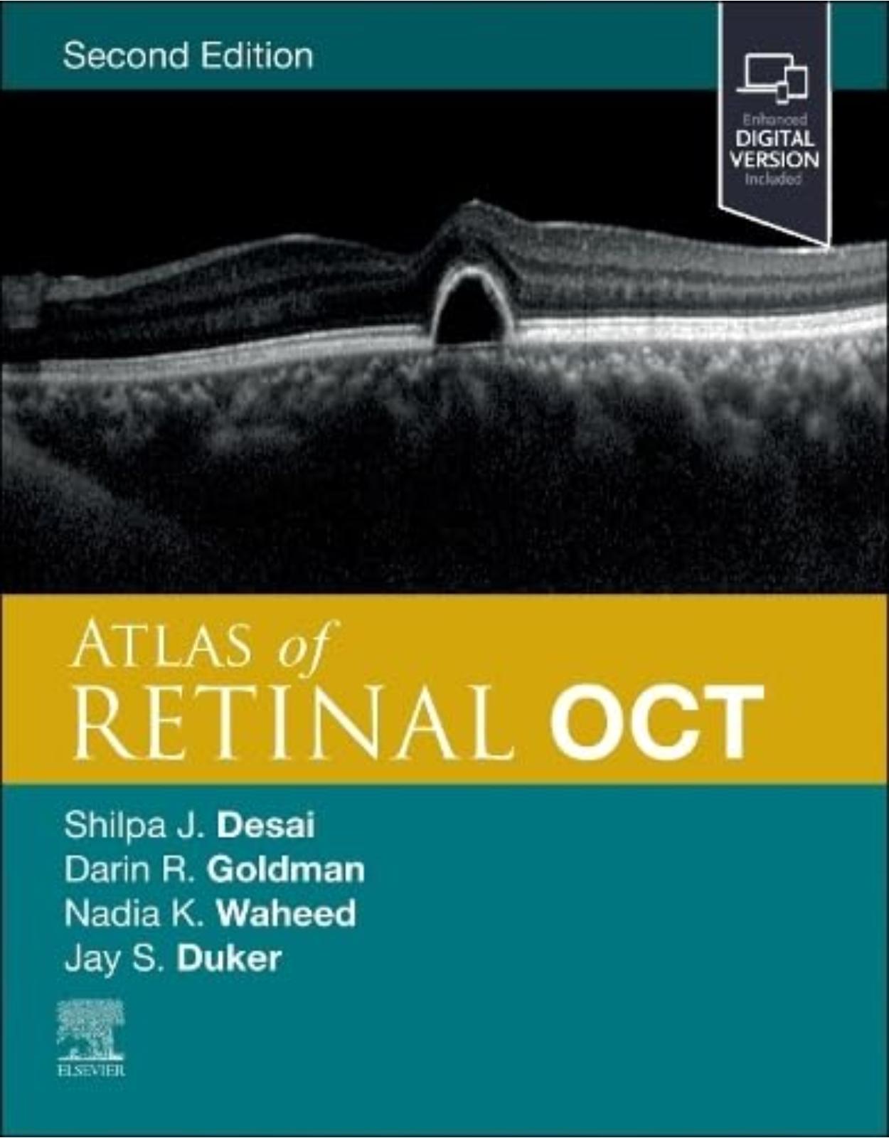
Atlas of Retinal OCT: Optical Coherence Tomography
Livrare gratis la comenzi peste 500 RON. Pentru celelalte comenzi livrarea este 20 RON.
Disponibilitate: La comanda in aproximativ 4-6 saptamani
Editura: Elsevier
Limba: Engleza
Nr. pagini: 272
Coperta: Hardcover
Dimensiuni: 216 x 276 mm
An aparitie: 3 Sept. 2023
|
Description: |
|
Features: |
|
Features more than 1,000 high-quality illustrations depicting the full spectrum of retinal diseases using OCT and OCTA scans, supported by clinical photos and ancillary imaging technologies. Contains new and updated image examples throughout—including new OCTA images with artifacts and key findings highlighted. Presents images as large as possible on the page with an abundance of arrows, pointers, and labels to guide you in pattern recognition and eliminate any uncertainty. Includes the latest high-resolution spectral domain OCT technology and new insights into OCT angiography technology to ensure you have the most up-to-date and highest quality examples available. Provides key feature points for each disorder, giving you the need-to-know OCT essentials for quick comprehension and rapid reference. An excellent diagnostic companion to Handbook of Retinal OCT: Optical Coherence Tomography, 2nd Edition. An digital version is included with purchase. The digital allows you to access all of the text, figures and references, with the ability to search, customize your content, make notes and highlights, and have content read aloud. |
Table of Contents:
Part 1: Normal Optical Coherence Tomography
Section 1: Normal Optic Nerve
1.1. Normal Optic Nerve
Summary
Volume Scans
Retinal Nerve Fiber Layer Thickness
Ganglion Cell Complex
Optic Nerve Morphology
Line Scans
References
Section 2: Normal Retina
2.1. Time Domain OCT
Summary
References
2.2. Spectral Domain OCT
Summary
Reference
2.3. Swept Source OCT
Summary
Section 3: Normal Choroid
3.1. Normal Choroid
Summary
References
Section 4: Normal Vitreous
4.1. Normal Vitreous
Summary
Key OCT Features
Reference
Section 5: OCT: Artifacts and Errors
5.1. OCT: Artifacts and Errors
Summary
Mirror Artifact
Vignetting
Misalignment
Software Breakdown
Blink Artifact
Motion Artifact
Out-of-Range Error
Reference
5.2. OCT Angiography Artifacts
Blockage Artifacts (Fig. 5.2.1)
White Line Artifacts (Fig. 5.2.2)
False Positive Flow
Quilting Defect (Fig. 5.2.3)
False Negative Flow
Projection Artifact (Fig. 5.2.4)
Vessel Suplication (Fig. 5.2.5)
Segmentation Errors (Fig. 5.2.6)
Shadowing Artifact (Fig. 5.2.7)
Wide-Field Artifacts (Fig. 5.2.8)
References
Part 2: Isolated Macular Disorders
Section 6: Age-Related Macular Degeneration
6.1.1. Drusen
Summary
Key Points
References
6.1.2. Geographic Atrophy
Summary
Key Points
References
6.1.3. Isolated Pigment Epithelial Detachment
Summary
Key Points
References
6.2.1. Type 1 Macular Neovascular Membrane
Summary
Key Points
References
6.2.2. Type 2 Macular Neovascular Membrane
Summary
Key Points
References
6.2.3. Type 3 Macular Neovascular Membrane
Summary
Key Points
Bibliography
6.2.4. Subretinal Hemorrhage
Summary
Key Points
Bibliography
6.2.5. Disciform Scar
Summary
Key Points
Bibliography
6.2.6. Retinal Pigment Epithelial Tear
Summary
Key Points
Bibliography
6.2.7. Polypoidal Choroidal Vasculopathy
Summary
Key Points
Bibliography
Section 7: Vitreomacular Interface Disorders
7.1. Vitreomacular Adhesion
Summary
Key OCT Features
7.2. Vitreomacular Traction
Summary
Key OCT Features
7.3. Full-Thickness Macular Hole
Summary
Key Points
Reference
7.4. Lamellar Macular Hole
Summary
Key OCT Features
Reference
7.5. Epiretinal Membrane
Summary
Key OCT Features
Reference
Section 8: Central Serous Chorioretinopathy
8.1. Central Serous Chorioretinopathy
Summary
OCT Key Findings
References
Section 9: Myopic Degenerative Maculopathies
9.1. Myopic Choroidal Neovascular Membrane
Summary
Key OCT Features
Reference
9.2. Myopic Macular Schisis
Summary
Key OCT Features
Reference
Bibliography
9.3. Dome-Shaped Macula
Summary
Key OCT Features
References
Bibliography
9.4. Posterior Staphyloma
Summary
Key OCT Features
References
Section 10: Hydroxycholoroquine and Pentosan Toxicities
10.1. Hydroxychloroquine Toxicity
Summary
References
10.2. Pentosan Toxicity
Summary
Key Features
References
Section 11: Vitelliform Macular Dystrophy
11.1. Vitelliform Dystrophy
Summary
Key Features
References
Section 12: Macular Telangiectasia
12.1. Macular Telangiectasia
Summary
Key Points
Section 13: Isolated Cystoid Macular Edema
13.1. Isolated Cystoid Macular Edema
Summary
Key Points
Bibliography
Section 14: Other Disorders Affecting the Macular
14.1. Angioid Streaks
Summary
Key Points
References
14.2. X-Linked Juvenile Retinoschisis
Summary
Key OCT Features
14.3. Oculocutaneous Albinism
Summary
Key OCT Finding
References
14.4. Subretinal Perfluorocarbon
Summary
Key OCT Features
Reference
Part 3: Vasoocclusive Disorders
Section 15: Diabetic Retinopathy
15.1. Diabetic Macular Edema
Summary
Key OCT Features
15.2. Nonproliferative Diabetic Retinopathy
Summary
Key OCT Features
15.3. Proliferative Diabetic Retinopathy
Summary
Key Points
Section 16: Retinal Venous Occlusive Disease
16.1. Branch Retinal Vein Occlusion
Summary
Key OCT Findings
Bibliography
16.2. Central Retinal Vein Occlusion
Summary
Key OCT Features
Bibliography
Section 17: Retinal Arterial Occlusive Disease
17.1. Branch Retinal Artery Occlusion
Summary
Key OCT Findings
Bibliography
17.2. Central Retinal Arterial Occlusion
Summary
Key OCT Findings
Bibliography
Part 4: Uveitis and Inflammatory Disorders
Section 18: Noninfectious Uveitis
18.1.1. Birdshot Retinochoroidopathy
Summary
Key OCT Features
Bibliography
18.1.2. Acute Posterior Multifocal Placoid Pigment Epitheliopathy
Summary
Key OCT Features
Bibliography
18.1.3. Multiple Evanescent White Dot Syndrome
Summary
Key OCT Features
Bibliography
18.1.4. Serpiginous Choroiditis
Summary
Key OCT Features
Bibliography
18.1.5. Multifocal Choroiditis and Panuveitis and Punctate Inner Choroidopathy
Summary
Key OCT Features
Reference
Bibliography
18.2. Vogt-Koyanagi-Harada Disease
Summary
Key OCT Features
Reference
Bibliography
18.3. Sympathetic Ophthalmia
Summary
Key OCT Findings
Bibliography
Section 19: Infectious Uveitis
19.1. Toxoplasmic Chorioretinitis
Summary
Key OCT Features
Bibliography
19.2. Acute Syphilitic Posterior Placoid Chorioretinitis
Summary
Key OCT Features
Bibliography
19.3. Tuberculosis
Summary
Key OCT Features
19.4. Posterior Scleritis
Summary
Key OCT Features
Bibliography
19.5. Candida Chorioretinitis
Summary
Key OCT Features
Bibliography
19.6. Acute Retinal Necrosis Syndrome
Summary
Key OCT Features
Bibliography
Part 5: Retinal and Choroidal Tumors
Section 20: Choroidal Tumors
20.1. Choroidal Nevus
Summary
Key OCT Features
Bibliography
20.2. Choroidal Melanoma
Summary
Key OCT Features
Bibliography
20.3. Solitary Choroidal Hemangioma
Summary
Key OCT Findings
References
Section 21: Retinal Tumors
21.1. Retinal Capillary Hemangioma
Summary
Key OCT Features
References
Section 22: Retinal Pigment Epithelium Tumors
22.1. Simple Hamartoma of the RPE
Summary
Key OCT Features
Reference
22.2. Combined Hamartoma of the Retina and RPE
Summary
Key OCT Features
Bibliography
Section 23: Metastatic Choroidal Tumors
23.1. Choroidal Metastases
Summary
Key OCT Features
References
Part 6: Trauma
Section 24: Mechanical Trauma
24.1. Valsalva Retinopathy
Summary
Key OCT Features
Bibliography
Section 25: Photic Maculopathy
25.1. Laser Maculopathy
Summary
Key OCT Features
Bibliography
25.2. Solar Maculopathy
Summary
Key OCT Features
Bibliography
Part 7: Inherited Retinal Degenerations
Section 26: Retinal Dystrophies
26.1. Retinitis Pigmentosa
Summary
Key Features
References
Bibliography
26.2. Stargardt Disease
Summary
Key OCT Features
26.3. Best Disease
Summary
Key OCT Features
26.4. Cone Dystrophy
Summary
Key Features
Reference
26.5. Malattia Leventinese (Doyne's Honeycomb Retinal Dystrophy)
Summary
Key OCT Features
Bibliography
26.6. Central Areolar Choroidal Dystrophy
Summary
Key OCT Features
References
Part 8: Vitreous Disorders
Section 27: Posterior Vitreous Detachment
27.1. Stages of Posterior Vitreous Detachment
Summary
Key OCT Features
Reference
Section 28: Asteroid Hyalosis
28.1. Asteroid Hyalosis
Summary
Key OCT Features
Section 29: Vitreous Hemorrhage
29.1. Vitreous Hemorrhage
Summary
Key OCT Features
Reference
Section 30: Vitreous Inflammation
30.1. Vitreous Inflammation
Summary
Key OCT Features
References
Part 9: Miscellaneous Retinal Disorders
Section 31: Peripheral Retinal Abnormalities
31.1. Tractional Retinal Detachment
Summary
Key OCT Features
31.2. Rhegmatogenous Retinal Detachment
Summary
Key OCT Features
References
31.3. Bullous Retinoschisis
Summary
Key OCT Features
Bibliography
31.4. Lattice Degeneration
Summary
Key OCT Features
Bibliography
31.5. Myelinated Nerve Fiber Layer
Summary
Key OCT Features
Index
| An aparitie | 3 Sept. 2023 |
| Autor | Jay S. Duker, Nadia K. Waheed, Darin Goldman, Shilpa J. Desai |
| Dimensiuni | 216 x 276 mm |
| Editura | Elsevier |
| Format | Hardcover |
| ISBN | 9780323930437 |
| Limba | Engleza |
| Nr pag | 272 |
| Versiune digitala | DA |
-
-
-
-
-
-
 1,18800 lei 1,05700 lei


Clientii ebookshop.ro nu au adaugat inca opinii pentru acest produs. Fii primul care adauga o parere, folosind formularul de mai jos.