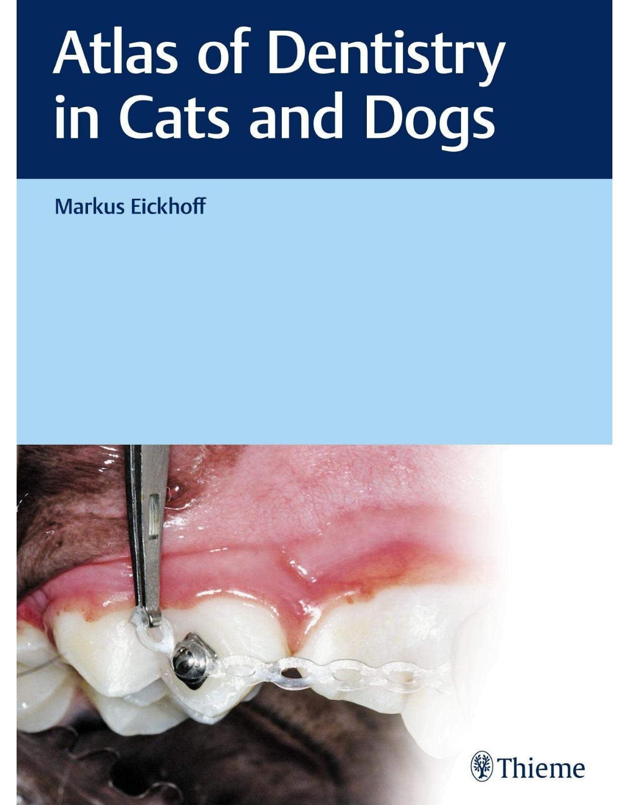
Atlas of Dentistry in Cats and Dogs
Livrare gratis la comenzi peste 500 RON. Pentru celelalte comenzi livrarea este 20 RON.
Disponibilitate: La comanda in aproximativ 4 saptamani
Autor: Markus Eickhoff
Editura: Thieme
Limba: Engleza
Nr. pagini: 468
Coperta: Hardcover
Dimensiuni: 30.99 x 23.11 cm
An aparitie: 30 May 2020
Description:
Dentistry at its finest
As a veterinarian and dentist in one, Dr. Markus Eickhoff has practiced high-level canine and feline dentistry for over 20 years. He now shares his fund of experience in this book with over 1500 images.
What equipment do I need? What treatment options are available and how do I perform them? This book provides detailed, systematic and generously illustrated instructions.
Over 1500 illustrations present details of many fascinating cases, allowing the practitioner to follow procedures in detail. The myriad practical tips and step-by-step instructions for performing common dental procedures provide security when performing dental treatment on small animals. Supported by the images, Dr. Eickhoff introduces the practitioner to commonly performed procedures such as the extraction of root fragments or the placement of fillings.
The focus on dogs and cats allows not only conventional treatment options but also less common ones to be presented in detail. Benefit from the latest insights for treating exceptional cases such as cleft palate or dental ankylosis.
Table of Contents:
Section I Fundamentals
1 Patient History
2 Examining the Head and Oral Cavity
2.1 Anatomy and Morphology of the Oral Cavity
2.2 Canine Oral Cavity
2.3 Feline Oral Cavity
2.4 Intraoral Radiography
2.5 Dental Probing
3 Interpreting Clinical Signs
3.1 Interpreting Clinical Signs of Pediatric Disorders
3.2 Interpreting Clinical Signs of Periodontal Disorders
3.3 Interpreting Clinical Signs of Trauma-related Conditions
3.4 Interpreting Clinical Signs of Resorptive Disorders
3.5 Interpreting the Clinical Signs of Mucosal Inflammatory Diseases
3.6 Interpreting Findings in Tumor Diseases
4 Treatment Aspects
4.1 Instruments and Equipment
4.1.1 Technical Resources
4.1.2 Instruments
4.2 Handling Instruments
4.3 Local Anesthesia
Section II Recurring Procedures
5 Dental Prophylaxis
5.1 Oral Hygiene Status
5.2 Dental Cleaning
5.2.1 Preparation
5.2.2 Ultrasonic Scaling
5.2.3 Dental Cleaning with Manual Tools
5.2.4 Polishing and Antiseptic Application
5.3 Toothbrushes
6 Tooth Extraction
6.1 Closed Extraction – Step by Step
6.1.1 Step 1
6.1.2 Step 2
6.1.3 Step 3
6.1.4 Step 4
6.2 Open Extraction of a Single-rooted Tooth – Step by Step
6.2.1 Step 1
6.2.2 Step 2
6.2.3 Step 3
6.2.4 Step 4
6.2.5 Step 5
6.2.6 Step 6
6.2.7 Step 7
6.3 Open Extraction of a Multirooted Tooth – Step by Step
6.3.1 Step 1
6.3.2 Step 2
6.3.3 Step 3
6.3.4 Step 4
6.3.5 Step 5
6.3.6 Step 6
6.3.7 Step 7
6.3.8 Step 8
6.3.9 Step 9
7 Retrieving Root Fragments
7.1 Retrieving Root Fragments – Step by Step
7.1.1 Step 1
7.1.2 Step 2
7.1.3 Step 3
7.1.4 Step 4
7.1.5 Step 5
7.1.6 Step 6
7.1.7 Step 7
7.1.8 Step 8
7.1.9 Step 9
8 Composite Fillings
8.1 Composite Fillings – Step by Step
8.1.1 Step 1
8.1.2 Step 2
8.1.3 Step 3
8.1.4 Step 4
8.1.5 Step 5
8.1.6 Step 6
9 Vital Pulpotomy
9.1 Crown Reduction – Step by Step
9.1.1 Step 1
9.1.2 Step 2
9.1.3 Step 3
9.1.4 Step 4
9.1.5 Step 5
9.1.6 Step 6
10 Crown Amputation
10.1 Crown Amputation – Step by Step
10.1.1 Step 1
10.1.2 Step 2
10.1.3 Step 3
10.1.4 Step 4
10.1.5 Step 5
11 Root Canal Fillings
11.1 Single-rooted Tooth – Step by Step
11.1.1 Step 1
11.1.2 Step 2
11.1.3 Step 3
11.1.4 Step 4
11.1.5 Step 5
11.1.6 Step 6
11.1.7 Step 7
11.1.8 Step 8
11.1.9 Step 9
11.1.10 Step 10
11.2 Multirooted Tooth 108 – Step by Step
11.2.1 Step 1
11.2.2 Step 2
11.2.3 Step 3
11.2.4 Step 4
11.2.5 Step 5
11.2.6 Step 6
11.2.7 Step 7
11.3 Multirooted Tooth 208 – Step by Step
11.3.1 Step 1
11.3.2 Step 2
11.3.3 Step 3
11.3.4 Step 4
11.3.5 Step 5
11.3.6 Step 6
12 Apicoectomy
12.1 Apicoectomy – Step by Step
12.1.1 Step 1
12.1.2 Step 2
12.1.3 Step 3
12.1.4 Step 4
12.1.5 Step 5
12.1.6 Step 6
13 Attaching Brackets
13.1 Attaching Brackets – Step by Step
13.1.1 Step 1
13.1.2 Step 2
13.1.3 Step 3
13.1.4 Step 4
13.1.5 Step 5
13.1.6 Step 6
14 Plate/Bite Plate
14.1 Acrylic Plate with Screw – Step by Step
14.1.1 Step 1
14.1.2 Step 2
14.1.3 Step 3
14.1.4 Step 4
14.1.5 Step 5
14.1.6 Step 6
14.1.7 Step 7
14.1.8 Step 8
Section III Case Studies
15 Young Animals
15.1 Missing Teeth
15.1.1 Multiple Missing Teeth (Hypodontia) and Reduced Tooth Development in Dogs
15.1.2 Hypodontia and Dental Fracture in Cats
15.1.3 Undeveloped Canines in Dogs
15.1.4 Retained Mandibular Premolar in a Dog and Severe Osteolysis
15.1.5 Bilateral Retained Mandibular Premolars in a Dog
15.1.6 Retained Maxillary Canine Tooth in a Dog
15.1.7 Orthodontic Treatment of a Retained Maxillary Canine Tooth in a Dog
15.2 Supernumerary Teeth
15.2.1 Persistent Deciduous Canines and Malpositioned Permanent Teeth in a Dog
15.2.2 Siblings with Hyperdontia
15.2.3 Double Maxillary Canines in a Cat
15.2.4 Malpositioned Incisors Due to Odontoma in a Dog
15.3 Dental Anomalies
15.3.1 Enamel Hypoplasia of Canines and Molars in a Dog
15.3.2 Generalized Enamel Hypoplasia with Root Deformity in a Dog
15.3.3 Dental Anomaly in a Dog
15.3.4 Double Crown of the Mandibular Premolars in a Cat
15.4 Malocclusions
15.4.1 Linguoversion and Mandibular Distoclusion
15.4.2 Rostral and Caudal Crossbite
15.4.3 Mesioverted Canine (Lance Canine)
15.5 Abrasions in a Young Animal
15.6 Tooth Fractures in Young Animals
15.6.1 Fractured Teeth 504 and 604.
15.6.2 Fractured Tooth 504
15.7 Persistent Deciduous Teeth
15.7.1 Persistent Deciduous Teeth
15.7.2 Shark Teeth in a Small Dog
15.8 Cleft Palate
15.8.1 Complete Cleft Palate in a Dog
15.8.2 Complete Cleft Palate Closure in a Dog, Two-stage Procedure
15.8.3 Trauma-induced Cleft Palate in a Cat
15.8.4 Cleft Lip and Palate in a Dog
15.9 Craniomandibular Osteopathy (CMO)
16 Teeth
16.1 Abrasion and Attrition
16.1.1 Severe Attrition of the Incisors
16.1.2 Severe Abrasion of the Front Teeth
16.1.3 Discoloration of the Maxillary Canine after Abrasion
16.1.4 Periapical Osteolysis of the Maxillary Carnassial Tooth after Abrasion
16.1.5 Abrasion of the Maxillary Canine with Fistula Formation
16.2 Tooth Fractures and Related Conditions
16.2.1 Tooth Discoloration
16.2.2 Tooth Fracture
16.2.3 Isolated Apical Process
16.2.4 Root Remnants
16.2.5 Vital Pulpotomy
16.2.6 Apexification
16.2.7 Bleaching
16.3 Caries
16.3.1 Caries on the Maxillary Cheek Teeth with Filling and Extraction
16.4 Fillings
16.4.1 Deformed Maxillary Canine Crown
16.4.2 Chipping of the Cusp and Buccal Surface on a Maxillary Carnassial Tooth
16.5 Crown Replacement
16.5.1 Metal Crown for Canine Tooth
16.5.2 Ceramic Canine Crown
16.5.3 Carnassial Tooth Crown
16.6 Feline Tooth Resorption
16.6.1 Schematics for Feline Tooth Resorption
16.6.2 Multiple Feline Resorptive Lesions
16.6.3 Tooth Resorption on Canine Roots
16.6.4 Development of Feline Tooth Resorption after Crown Amputation
16.7 Canine Tooth Resorption
16.7.1 Canine Tooth Resorption on Tooth 309
16.8 Tooth Displacement
16.8.1 Displacement of the Maxillary Left Canine
16.8.2 Avulsion of the Maxillary Right Canine Tooth
16.9 Tooth Extraction
16.9.1 Open Extraction of the Maxillary Canine Tooth in a Dog
16.9.2 Open Extraction of the Maxillary Canine Tooth in a Cat
16.9.3 Extraction of Multiple Maxillary Cheek Teeth in a Cat
16.9.4 Extraction of a Root Fragment of a Maxillary Carnassial Tooth in a Cat
16.9.5 Extraction of Teeth with Root Resorption in a Dog
16.10 Dental Implant of a Canine Tooth
17 Periodontium
17.1 Periodontium: Physiology and Pathology
17.1.1 Evaluating the Periodontium in a Dog
17.1.2 Evaluating the Periodontium in a Cats
17.2 Periodontitis
17.2.1 Gingivitis in the Dog
17.2.2 Effect of Dental Cleaning on the Canine Gingiva
17.2.3 Gingivectomy in a Cat with Gingival Hyperplasia
17.2.4 Generalized Periodontitis in a Dog
17.2.5 Generalized Periodontitis in a Cat
17.2.6 Fistula Formation in Association with Periodontitis
17.2.7 Symmetrical Advanced Periodontitis at the Maxillary Cheek Teeth
17.2.8 Local Interdental Periodontitis
17.2.9 Labial Gingivoplasty in a Dog with Local Periodontitis
17.2.10 Malocclusion-induced Local Periodontitis
17.2.11 Local Periodontitis due to a Crossbite of the Front Teeth
17.2.12 Local Periodontitis and Kissing Ulcers
17.2.13 Lasers
17.2.14 Laser Applications in Periodontology
17.2.15 Laser Gingivectomy in Cats
17.2.16 Dissecting Periodontal Mucosa
17.2.17 Covering Gingival Recession at the Maxillary Carnassial Tooth in a Cat
17.2.18 Guided Tissue Regeneration and Guided Bone Regeneration
17.3 Gingival Hyperplasia
17.3.1 Gingival Hyperplasia and Gingivectomy
17.3.2 Gingival Hyperplasia and Pseudopockets
17.3.3 Gingival Hyperplasia in a Cat
17.3.4 Feline Gingival Hyperplasia and Extractions
17.4 Oronasal Fistula
17.4.1 Symmetrical Oronasal Fistulas of the Maxillary Canine Teeth
17.4.2 Closing an Oronasal Fistula at the Maxillary Left Canine Tooth
17.5 Gingivostomatitis
17.5.1 Gingivostomatitis with Extraction of All Cheek Teeth
17.5.2 Gingivostomatitis in a Young Cat
17.5.3 Delayed Recovery from Gingivostomatitis
17.5.4 Gingivostomatitis Before and After Extraction of All Teeth
17.6 Stomatitis in a Dog
17.6.1 Polypoid Stomatitis
17.6.2 Mucositis
18 Oral Mucosa
18.1 Immunogenic Inflammation
18.1.1 Contact Ulcer
18.1.2 Eosinophilic Granuloma Complex
18.1.3 Systemic Lupus Erythematosus (SLE)
18.1.4 Eosinophilic Myositis
18.1.5 Lip Fold Dermatitis
18.2 Trauma
18.2.1 Stick Injury to the Palate
18.2.2 Fistula Formation after a Stick Injury to the Palate
18.2.3 Avulsion of the Skin over the Mandible after an Accident
19 Oral Masses
19.1 Cysts
19.1.1 Follicular Cyst in the Mandible Near a Partially Retained Premolar
19.1.2 Symmetrical Follicular Cysts in the Mandible
19.1.3 Symmetrical Ranula Formation
19.2 Tumors
19.2.1 Squamous Cell Carcinoma at the Mandibular Front Teeth of a Dog
19.2.2 Squamous Cell Carcinoma in the Maxilla of a Cat
19.2.3 Squamous Cell Carcinoma in the Mandible of a Cat
19.2.4 Diagram of Jaw Resection
19.2.5 Acanthomatous Ameloblastoma at the Caudal Body of the Mandible in a Dog
19.2.6 Acanthomatous Ameloblastoma at the Mandibular Front Teeth of a Dog
19.2.7 Papilloma in a Young Dog
19.2.8 Odontoma in a Dog
19.2.9 Odontoma in a Cat
19.2.10 Symmetrical Tissue Granulation in the Mandible of a Cat
19.2.11 Treatment of Inflammatory Oral Masses through Crown Reduction of the Carnassial Teeth in a Cat
19.2.12 Other Diagnostic Imaging
20 Jawbone
20.1 Jaw Fractures
20.1.1 Noninvasive Repair of a Fractured Body of the Mandible in a Dog
20.1.2 Nasal Fracture
20.1.3 Nasal Avulsion
20.1.4 Symphyseal Separation in a Cat
20.1.5 Fracture of the Caudal Body of the Mandible in a Cat
20.1.6 TMJ Fracture in a Cat
20.1.7 Carnassial Tooth in the Fracture Gap
20.2 TMJ Luxation
20.2.1 TMJ Luxation in a Dog
20.2.2 TMJ Luxation in a Cat
20.3 TMJ Dysplasia
20.3.1 TMJ Dysplasia in a Dog
20.3.2 TMJ Dysplasia in a Cat
Section IV Appendix
21 Selected References
About the Authors
Contact Information
Index
Imprint
| An aparitie | 30 May 2020 |
| Autor | Markus Eickhoff |
| Dimensiuni | 30.99 x 23.11 cm |
| Editura | Thieme |
| Format | Hardcover |
| ISBN | 9783132432826 |
| Limba | Engleza |
| Nr pag | 468 |
Clientii ebookshop.ro nu au adaugat inca opinii pentru acest produs. Fii primul care adauga o parere, folosind formularul de mai jos.