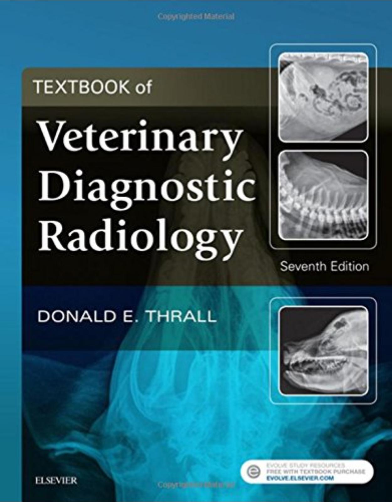
Textbook of Veterinary Diagnostic Radiology
Livrare gratis la comenzi peste 500 RON. Pentru celelalte comenzi livrarea este 20 RON.
Disponibilitate: La comanda in aproximativ 4 saptamani
Editura: Elsevier
Limba: Engleza
Nr. pagini: 1000
Coperta: Hardcover
Dimensiuni: 28.4 x 22.6 x 5 cm
An aparitie: 2 Feb 2018
|
Description: Learn the latest advances in veterinary diagnostic radiology! Textbook of Veterinary Diagnostic Radiology, 7th Edition, is a one-stop resource covering the principles of radiographic technique and interpretation for dogs, cats, and horses. Within this bestselling text, high-quality radiographic images accompany clear coverage of diagnostic radiology, ultrasound, MRI, and CT. User-friendly direction helps you to develop essential skills in patient positioning, radiographic technique and safety measures, normal and abnormal anatomy, radiographic viewing and interpretation, and alternative imaging modalities. This new edition has been thoroughly revised to include important advances in the field, information about contrast media, dental radiography, and more! |
||
|
Features: |
||
|
Coverage of ultrasound imaging procedures such as the esophagram, upper GI examination, excretory urography, and cystography helps in determining when and how these procedures are performed in today’s practice. Rewritten chapters on basic interpretation emphasizes radiography, radiation safety, superficial coverage of normal variants, and will include more in-depth information on the framework for interpretation. An atlas of normal radiographic anatomy in each section makes it easier to recognize abnormal radiographic findings. High-quality radiographic images clarify key concepts and interpretation principles. Up-to-date coverage of the most commonly seen species in private veterinary practices and veterinary teaching hospitals includes the cat, dog, and horse. |
||
|
New To This Edition: |
||
|
NEW! Chapter on CT and MR contrast media gives you a better understanding of the agents used to alter patient contrast. NEW! Information on digital imaging helps you understand the latest advances in digital imaging. NEW! Chapter on dental radiology covers common dental issues you may encounter in practice. NEW! Chapter on MR spinal imaging provides the latest information on the diagnosis of spinal cord disease through the use of CT and MRI. |
||
|
Table Of Contents: |
||
|
Section I: Physics and Principles of Interpretation
Section II: The Axial Skeleton: Canine, Feline, and Equine
Section III: The Appendicular Skeleton: Canine, Feline, and Equine
Section IV: Thoracic Cavity: Canine, Feline, and Equine
Section V: Abdominal Cavity: Canine and Feline
|
||
| An aparitie | 2 Feb 2018 |
| Autor | Donald E. Thrall DVM PhD DACVR |
| Dimensiuni | 28.4 x 22.6 x 5 cm |
| Editura | Elsevier |
| Format | Hardcover |
| ISBN | 9780323482479 |
| Limba | Engleza |
| Nr pag | 1000 |
-
 1,06400 lei 92000 lei
 -

-


Clientii ebookshop.ro nu au adaugat inca opinii pentru acest produs. Fii primul care adauga o parere, folosind formularul de mai jos.