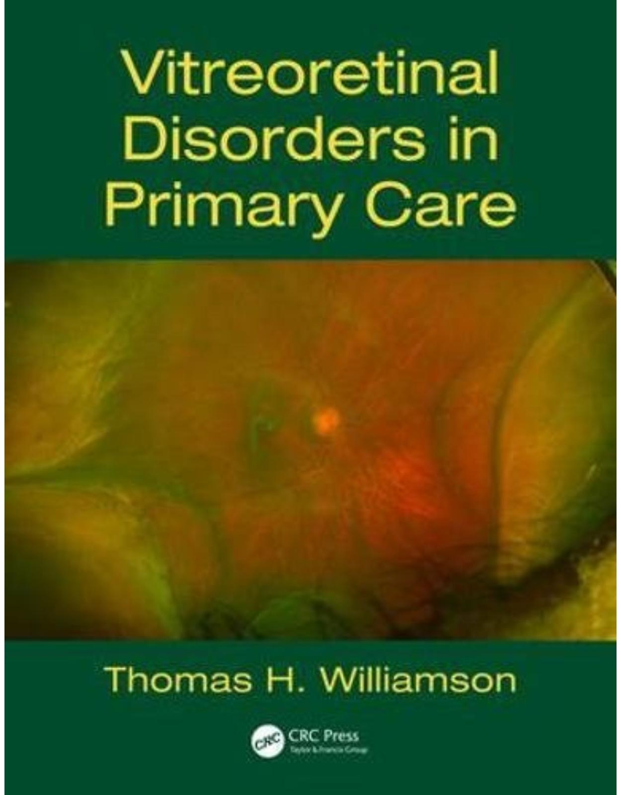
Vitreoretinal Disorders in Primary Care
Livrare gratis la comenzi peste 500 RON. Pentru celelalte comenzi livrarea este 20 RON.
Disponibilitate: La comanda in aproximativ 4 saptamani
Autor: Thomas H. Williamson
Editura: CRC Press
Limba: Engleza
Nr. pagini: 242
Coperta: Paperback
Dimensiuni: 19.05 x 1.27 x 24.77 cm
An aparitie: 23 Oct. 2017
Description:
Emergency ophthalmology is an area full of pitfalls for the unwary primary care practitioner. Vitreoretinal disorders make up the majority of emergency sight-threatening conditions, and a wide and increasingly varied range of conditions of the eye present in primary care settings. Correct diagnosis at initial presentation, and appropriate and speedy referral, are extremely important. This book is therefore an essential reference for the primary care physician, who is often the first to see these patients and is in a position of responsibility for decision-making.
Table of Contents:
Chapter 1 Anatomy and examination of the eye
Embryology of the eye
Anatomy
Vitreous
Anatomical Attachments of the Vitreous to the Surrounding Structures
Retina
Retinal Pigment Epithelium
Photoreceptor Layer
Cones
Ganglion Cells
Nerve Fibre Layer
Inner Limiting Membrane
Retinal Blood Vessels
Bruch’s Membrane
Choroid
Investigation
Visual Acuity
Slit Lamp
Optical Coherence Tomography
Inner Segment and Outer Segment Junction (Ellipsoid Layer)
Central Retinal Thickness
Subjective Tests
References
Chapter 2 Posterior vitreous detachment
Introduction
Symptoms
Floaters
Flashes
Signs
Detection of PVD
Shafer’s Sign
Vitreous Haemorrhage
Ophthalmoscopy
Retinal tears
U tears
Atrophic Round Holes
Other Breaks
Progression to Retinal Detachment
Peripheral retinal degenerations
Referral
Posterior Vitreous Detachment
Medicolegal Case
Missing the Symptoms of Posterior Vitreous Detachment
Error
Errors
References
Chapter 3 Vitreous haemorrhage
Introduction
Aetiology
Causes4–10
Natural History
Erythroclastic Glaucoma
Investigation
Ultrasound
Ultrasound Features
Referral
Vitreous Haemorrhage Non-Diabetic
Medicolegal Case
References
Chapter 4 Rhegmatogenous retinal detachment
Introduction
Tears with Posterior Vitreous Detachment
Breaks Without Posterior Vitreous Detachment
Natural History
Chronic RRD
Clinical Features
Anterior Segment Signs
Signs in the Vitreous
Subretinal Fluid Accumulation
Retinal Break Patterns in RRD
Macula Off or On
Flat Retinal Breaks
Retinopexy
Cryotherapy
Laser
Rhegmatogenous Retinal Detachment
Principles of Surgery
Pars Plana Vitrectomy
Posturing
Non-Drain Procedure
Pneumatic Retinopexy
Success Rates
Causes of Failure
Proliferative Vitreoretinopathy
Introduction
Pathogenesis
Clinical Features
Introduction
Grading
Risk of PVR
Surgery
Relieving Retinectomy
Success Rates
Surgery for Redetachment
Secondary Macular Holes
Detachment with Choroidal Effusions
Medicolegal Cases
Case 1
Errors
Case 2
Errors
Case 3
Errors
References
Chapter 5 Different presentations of rhegmatogenous retinal detachments
Age-related RRD from PVD
Atrophic hole RRD with attached vitreous
Pseudophakic RRD
Aphakic RRD
Retinal dialysis
Clinical features
Giant retinal dialysis
Par ciliaris tear
Giant retinal tear
Clinical features
Stickler’s syndrome
Other eye
Retinal detachment in high myopes
Clinical features
Retinoschisis-related retinal detachment
Clinical features
Infantile retinoschisis
Senile retinoschisis
Differentiation of retinoschisis from chronic RRD
Retinal detachment in retinoschisis
Juvenile retinal detachment
Atopic dermatitis
Refractive surgery
Congenital cataract
Others
References
Chapter 6 Macular disorders
Introduction
Idiopathic macular hole
Clinical features
Introduction
Watzke–Allen test
Grading
Natural history
Optical coherence tomography
Secondary macular holes
Lamellar and partial thickness holes
Pars plana vitrectomy
Microplasmin
Referral
Macular pucker and vitreomacular traction
Clinical features
Other conditions
Secondary Macular Pucker
Success rates of surgery
Specific complications of surgery
Membrane recurrence
Referral
Age-related macular degeneration
Clinical features
Simplified AREDS scoring system
Vitreous haemorrhage and choroidal neovascular membranes
Referral
Pneumatic displacement of subretinal haemorrhage
Referral
Choroidal neovascular membrane not from AMD
Introduction
References
Chapter 7 Diabetic retinopathy
Introduction
Diabetic retinopathy
Introduction
Diabetic retinopathy grading
Diabetic vitreous haemorrhage
Clinical features
Diabetic retinal detachment
Clinical features
Success rates
Diabetic maculopathy
Retinal vein occlusion
Sickle cell disease
Introduction
Types of sickle cell disease
Systemic investigation
Inheritance and race
Systemic manifestations
Ophthalmic presentation
Visual outcome
Screening
Survival
Retinal vasculitis
Central retinal artery occlusion
Medicolegal case
References
Chapter 8 Trauma
Introduction
Classification
Contusion injuries
Clinical presentation
Types of retinal break
Dialysis
Par ciliaris tears
Ragged tear in commotio retinae
Giant retinal tears
Visual outcome
Rupture
Clinical presentation
Visual outcome
Penetrating injury
Clinical presentation
Endophthalmitis
Retinal detachment
Visual outcome
Trauma scores
Intraocular foreign bodies
Clinical presentation
Diagnostic imaging
IOFB materials
Visual outcome
Perforating injury
Sympathetic ophthalmia
Proliferative vitreoretinopathy
Phthisis bulbi
Referral
Medicolegal case
Case 1
Case 2
References
Chapter 9 Complications of anterior segment surgery
Introduction
Dropped nucleus
Clinical features
Success rates
Intraocular lens dislocations
Clinical presentation
Post-operative endophthalmitis
Clinical features
Infective organisms
Antibiotics
Success rates
Chronic post-operative endophthalmitis
Needle-stick injury
Clinical features
Intraocular haemorrhage
Retinal detachment
Chronic uveitis
Post-operative vitreomacular traction
Post-operative choroidal effusion
Medicolegal case
References
Chapter 10 Uveitis and allied disorders
Non-infectious uveitis of the posterior segment
Referral for Vitrectomy
Vitreous Opacification
Retinal Detachment
Cystoid Macular Oedema
Hypotony
Infectious Uveitis
Acute Retinal Necrosis
Clinical Features
Cytomegalovirus retinitis
Clinical Features
Fungal endophthalmitis
Clinical Features
Other infections
Ocular Lymphoma
Clinical Features
Visual Outcome and Survival
Paraneoplastic Retinopathy
References
Chapter 11 Miscellaneous conditions
Vitrectomy for Vitreous Opacities
Vitreous Anomalies
Persistent Hyperplastic Primary Vitreous
Asteroid Hyalosis
Amyloidosis
Retinal Haemangioma and Telangiectasia
Optic Disc Anomalies
Optic Disc Pits and Optic Disc Coloboma
Morning Glory Syndrome
Retinochoroidal Coloboma
Marfan’s Syndrome
Retinopathy of Prematurity
Uveal Effusion Syndrome
Clinical Features
Terson’s Syndrome
Disseminated Intravascular Coagulation
Retinal Prosthesis
References
Index
Read Less
| An aparitie | 23 Oct. 2017 |
| Autor | Thomas H. Williamson |
| Dimensiuni | 19.05 x 1.27 x 24.77 cm |
| Editura | CRC Press |
| Format | Paperback |
| ISBN | 9781138628113 |
| Limba | Engleza |
| Nr pag | 242 |


Clientii ebookshop.ro nu au adaugat inca opinii pentru acest produs. Fii primul care adauga o parere, folosind formularul de mai jos.