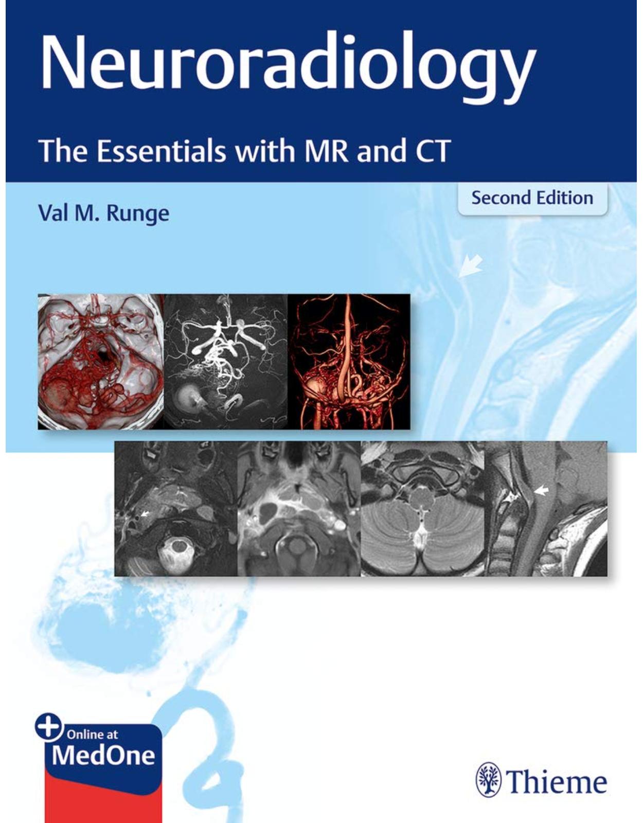
Neuroradiology: The Essentials with MR and CT
Livrare gratis la comenzi peste 500 RON. Pentru celelalte comenzi livrarea este 20 RON.
Disponibilitate: La comanda in aproximativ 4 saptamani
Autor: Val M. Runge
Editura: Thieme
Limba: Engleza
Nr. pagini: 254
Coperta: Paperback
Dimensiuni: 27.94 x 21.59 cm
An aparitie: 1 Sept. 2020
Description:
An image-rich neuroradiology reference and board prep from renowned experts
Neuroradiology: The Essentials with MR and CT, Second Edition, written by world-renowned neuroradiologist and MRI pioneer Val Runge, builds on the acclaimed prior edition. The splendidly illustrated compendium features in-depth discussion of important imaging findings, focused primarily on common disease processes. An impressive cadre of international experts contribute to the text, which is written from a clinical radiology perspective and draws from firsthand experiences. MRI physics pearls and tips throughout the book will help radiologists avoid common pitfalls.
Designed as a practical educational resource for clinical neuroradiology, the text is divided into three sections: the brain, head and neck, and spine. The brain and spine chapters are divided into subsections covering normal anatomy and major disease categories such as congenital, traumatic, degenerative, vascular, infectious, and neoplastic. Head and neck chapters are organized by major anatomic region. Clinical cases encompass the use of advanced imaging techniques such as perfusion, high-resolution imaging, and spectroscopy.
Key Features
About 1,300 high-quality MR and CT images illustrate relevant findings and cases, including those often not well-described in more traditional academic textbooks
New figures, updates on ultra-high-field 7T MRI, and additional in-depth text on cerebrovascular disease – especially brain aneurysms and AVMs
Covers a wide array of diseases – from stroke and multiple sclerosis to cases one might see once a year, such as glutaric acidemia type 1 and CADASIL
This excellent clinical resource provides a robust study prep for the boards and is a must-read for radiology residents prior to neuroradiology rotation. A quick reference for diagnosing challenging cases encountered in daily practice, it will also benefit neuroradiology fellows and general radiologists.
Table of Contents:
1. Brain
1.1 Normal Anatomy and Common Variants.
1.1.1 Normal Intracranial Anatomy
1.1.2 Normal Arterial Anatomy
1.1.3 Normal Venous Anatomy
1.1.4 Normal Myelination
1.1.5 Variants Involving the Septum Pellucidum
1.1.6 Physiological Calcification
1.1.7 Incidental Cystic Lesions
1.1.8 Dilated Perivascular Spaces
1.1.9 Other Incidental Lesions
1.2 Congenital Malformations
1.2.1 Posterior Fossa Malformations
1.2.2 Cortical Malformations
1.2.3 Callosal Malformations
1.2.4 Holoprosencephaly and Related Disorders
1.2.5 Phakomatoses
1.2.6 Lipomas
1.2.7 Anomalies of
1.3 Inherited Metabolic Disorders
1.3.1 Diseases Affecting White Matter
1.3.2 Disease Affecting Gray Matter: Huntington Disease
1.3.3 Diseases Affecting Both White and Gray Matter
1.4 Acquired Metabolic, Systemic, and Toxic Disorders Toxic Disorders
1.4.1 Acute Hypertensive Encephalopathy
1.4.2 Wernicke Encephalopathy
1.4.3 Hepatic Encephalopathy
1.4.4 Carbon Monoxide Poisoning
1.4.5 Osmotic Demyelination
1.4.6 Mesial Temporal Sclerosis
1.5 Hemorrhage
1.5.1 Parenchymal Hemorrhage
1.5.2 Subarachnoid Hemorrhage
1.5.3 Superficial Siderosis
1.6 Trauma
1.6.1 Parenchymal Injury
1.6.2 Epidural Hematoma
1.6.3 Subdural Hematoma
1.6.4 Nonaccidental Trauma (Child Abuse)
1.6.5 Penetrating Injuries
1.7 Herniation
1.8 Infarction
1.8.1 Arterial Territory Infarcts
1.8.2 Lacunar Infarcts
1.8.3 Medullary Infarcts
1.8.4 Temporal Evolution
1.8.5 Abnormal Contrast Enhancement
1.8.6 CT in Infarction
1.8.7 Chronic Infarcts
1.8.8 Hemorrhagic Transformation
1.8.9 Periventricular Leukomalacia
1.9 Dementia and Degenerative Disease
1.9.1 Alzheimer Disease
1.9.2 Frontotemporal Dementia
1.9.3 Multisystem Atrophy
1.9.4 Small Vessel White Matter Ischemic Disease
1.10 Vasculitis and Vasculitides
1.10.1 Sickle Cell Disease
1.10.2 Moyamoya Disease
1.10.3 CADASIL and Behçet Disease .
1.10.4 Systemic Lupus Erythematosus
1.11 Vascular Lesions
1.11.1 Aneurysms
1.11.2 Vascular Malformations
1.11.3 Sinus Thrombosis
1.12 Infection and Inflammation
1.12.1 Parenchymal Abscess
1.12.2 Epidural and Subdural Abscesses
1.12.3 Meningitis
1.12.4 Ventriculitis
1.12.5 Encephalitis
1.12.6 Toxoplasmosis
1.12.7 Neurocysticercosis
1.12.8 Tuberculosis
1.12.9 Creutzfeldt-Jakob Disease
1.12.10 Neurosarcoidosis
1.12.11 HIV/AIDS
1.13 Demyelinating Disease
1.13.1 Multiple Sclerosis
1.13.2 Neuromyelitis Optica
1.13.3 Acute Disseminated Encephalomyelitis
1.14 Neoplasms
1.14.1 Pilocytic Astrocytoma
1.14.2 Low-grade Astrocytoma
1.14.3 Anaplastic Astrocytoma
1.14.4 Glioblastoma Multiforme
1.14.5 Gliomatosis Cerebri
1.14.6 Oligodendroglioma
1.14.7 Ganglioglioma
1.14.8 Hemangioblastoma
1.14.9 Primary CNS Lymphoma
1.14.10 Medulloblastoma
1.14.11 Supratentorial PNET
1.14.12 Dysembryoplastic Neuroepithelial Tumor
1.14.13 Choroid Plexus Papilloma
1.14.14 Ependymoma
1.14.15 Pituitary Microadenoma
1.14.16 Pituitary Macroadenoma
1.14.17 Craniopharyngioma
1.14.18 Pineal Region Neoplasms
1.14.19 Brain (Parenchymal) Metastases
1.14.20 Leptomeningeal Metastases
1.14.21 Calvarial Metastases
1.14.22 Langerhans Cell Histiocytosis
1.14.23 Calvarial Hemangioma
1.14.24 Fibrous Dysplasia
1.14.25 Meningioma
1.14.26 Hemangiopericytoma
1.14.27 Radiation Injury
1.14.28 Radiation Necrosis
1.15 Nonneoplastic Cysts
1.15.1 Arachnoid Cyst
1.15.2 Epidermoid Cyst
1.15.3 Dermoid Cyst
1.15.4 Colloid Cyst
1.16 Cerebrospinal Fluid Disorders
1.16.1 Obstructive Hydrocephalus, Intraventricular
1.16.2 Obstructive Hydrocephalus, Extraventricular
1.16.3 Normal Pressure Hydrocephalus
1.16.4 CSF Shunts and Complications
1.16.5 Idiopathic Intracranial Hypertension
1.16.6 Intracranial Hypotension
2. Head and Neck
2.1 Skull Base
2.2 Temporal Bone
2.2.1 Neoplasms
2.3 Orbit
2.3.1 Inflammation/Infection
2.3.2 Neoplasms
2.4 Globe
2.5 Visual Pathway
2.6 Paranasal Sinuses, Nasal Cavity, and Face
2.6.1 Inflammation/Infection
2.6.2 Fractures
2.6.3 Sinus Surgery
2.6.4 Neoplasms
2.7 Mandible
2.8 Temporomandibular Joint
2.9 Nasopharynx
2.10 Oral Cavity, Oropharynx
2.11 Salivary Glands
2.12 Parapharyngeal Space
2.13 Larynx
2.14 Soft Tissues of the Neck
2.14.1 Lymph Nodes
2.14.2 Congenital Anomalies
2.14.3 Inflammation/Infection
2.14.4 Neoplasms
2.14.5 Vascular Lesions
3. Spine
3.1 Normal Anatomy, Imaging Technique, and Common Variants
3.1.1 Anatomy of the Normal Spine
3.1.2 Imaging Technique
3.1.3 Common Normal Variants and Incidental Findings
3.1.4 Incidental Cystic Lesions
3.2 Congenital Disease
3.2.1 Congenital Spinal Stenosis
3.2.2 Scoliosis
3.2.3 Tethered Cord
3.2.4 Syringohydromyelia
3.2.5 Meningomyeloceles and Lipomyelomeningoceles
3.2.6 Diastematomyelia
3.2.7 Caudal Regression
3.2.8 Anterior Sacral Meningocele
3.2.9 Dorsal Dermal Sinus
3.2.10 Intraspinal Enteric Cyst (Neurenteric Cyst)
3.2.11 Spinal Cord Herniation
3.2.12 Dermoid and Epidermoid Cysts
3.2.13 Neurofibromatosis
3.2.14 Klippel-Feil
3.2.15 Achondroplasia
3.3 Trauma
3.3.1 Cervical Spine Trauma
3.3.2 Burst Fracture
3.3.3 Flexion Injury
3.3.4 Benign Osteoporotic Fractures
3.3.5 Spinal Cord Injury
3.3.6 Epidural Hemorrhage
3.3.7 Brachial Plexus Injury
3.4 Degenerative Disease
3.4.1 Degenerative Spinal Stenosis
3.4.2 Disk, Endplate, Foraminal, and Spinal Canal Disease
3.4.3 Abnormalities of Vertebral Alignment
3.4.4 Surgery
3.4.5 Spondyloarthropathies
3.5 Arteriovascular Disease and Ischemia
3.5.1 Spinal Dural Arteriovenous Fistulas
3.5.2 Spinal Cord Arteriovenous Malformations
3.5.3 Spinal Cord Arterial Ischemia
3.5.4 Cavernous Malformation
3.6 Infection and Inflammation
3.6.1 Disk Space Infection
3.6.2 Tuberculous Spondylitis
3.6.3 Epidural Abscess
3.6.4 Meningitis and Myelitis
3.6.5 Arachnoiditis
3.6.6 Guillain-Barré
3.6.7 Chronic Inflammatory Demyelinating Polyneuropathy
3.6.8 Sarcoidosis
3.6.9 Multiple Sclerosis
3.6.10 Neuromyelitis Optica
3.6.11 Acute Transverse Myelitis
3.6.12 Vitamin B12Deficiency
3.6.13 Paget Disease
3.7 Neoplasms
3.7.1 Nerve Sheath Tumors (Neurofibroma, Schwannoma)
3.7.2 Meningioma
3.7.3 Ependymoma
3.7.4 Astrocytoma
3.7.5 Hemangioblastoma
3.7.6 Leptomeningeal and Spinal Cord Metastases
3.7.7 Vertebral Body Hemangioma
3.7.8 Aneurysmal Bone Cyst
3.7.9 Osteoid Osteoma
3.7.10 Osteochondroma
3.7.11 Giant Cell Tumor
3.7.12 Chordoma
3.7.13 Sacrococcygeal Teratoma
3.7.14 Focal Vertebral Body Metastatic Disease
3.7.15 Pathologic Compression Fracture
3.7.16 Langerhans Cell Histiocytosis
3.7.17 Diffuse Marrow Disease
3.7.18 Lymphoma
3.7.19 Leukemia
3.7.20 Multiple Myeloma
Index
| An aparitie | 1 Sept. 2020 |
| Autor | Val M. Runge |
| Dimensiuni | 27.94 x 21.59 cm |
| Editura | Thieme |
| Format | Paperback |
| ISBN | 9781684201532 |
| Limba | Engleza |
| Nr pag | 254 |







Clientii ebookshop.ro nu au adaugat inca opinii pentru acest produs. Fii primul care adauga o parere, folosind formularul de mai jos.