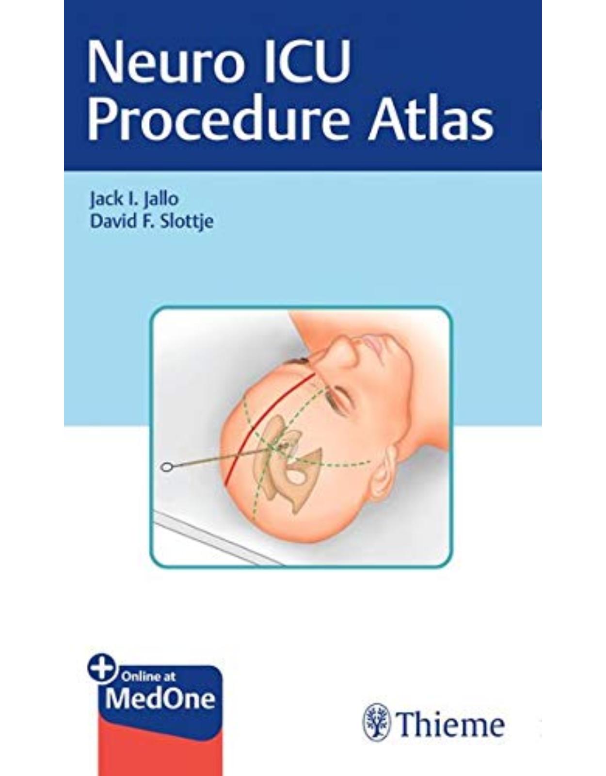
Neuro ICU Procedure Atlas
Livrare gratis la comenzi peste 500 RON. Pentru celelalte comenzi livrarea este 20 RON.
Disponibilitate: La comanda in aproximativ 4-6 saptamani
Autor: Jack I. Jallo, David Slottje
Editura: Thieme
Limba: Engleza
Nr. pagini: 174
Coperta: Paperback
Dimensiuni: 20.32 x 12.7 cm
An aparitie: 25 Jan. 2021
Description:
Atlas provides insightful guidance on how to perform core procedures in the neuro ICU
Neuro ICU Procedure Atlas by distinguished neurosurgeons Jack I. Jallo and David F. Slottje is a visually rich, step-by-step atlas describing technical aspects of common bedside procedures performed in patients with grave neurological conditions. With contributions from an impressive group of contributors, the book features 13 chapters encompassing fundamental techniques, such as shunt externalization/shunt tap, lumbar puncture, lumbar drain, ICP monitor, PbtO2 and JvO2 monitors, central and arterial line, cervical traction, intubation, cricothyrotomy, chest tube, and more.
Key Highlights:
In-depth, reader friendly discussion of vital clinical issues including indications and contraindications, relevant anatomy/physiology, and complications
Clinical pearls and troubleshooting sections for each approach
Illustrations of ventricular drain or ICP monitor placement, non-conventional ventricular drain insertion points, trajectory lines for ventricular drain insertion, and more provide detailed visual learning guides
This essential resource will help neurosurgical residents and early-career neurosurgeons, intensivists, nurse practitioners, and physician assistants accelerate the learning curve for the bedside care of critically ill patients.
Table of Contents:
1 External Ventricular Drain
1.1 Introduction
1.2 Relevant Anatomy and Physiology
1.3 Indications
1.3.1 ICP Monitoring
1.3.2 CSF Diversion
1.3.3 Intrathecal Access
1.4 Contraindications
1.4.1 Special Situations
1.5 Equipment
1.6 Technique
1.6.1 Preparation
1.6.2 Medications
1.6.3 Positioning/Equipment Setup
1.6.4 Procedure
1.6.5 Alternative Approaches
1.7 Complications
1.7.1 Infection
1.7.2 Hemorrhage
1.7.3 Upward Herniation
1.7.4 Aneurysm Re-rupture
1.7.5 Motor Cortex Injury
1.7.6 Superior Sagittal Sinus Injury
1.8 Expert Suggestions/Troubleshooting
1.8.1 Scalp Bleeding
1.8.2 Entry Site
1.8.3 Tunneling Direction
1.8.4 Fracture Lines/Cranial Defects
1.8.5 Ventricular Collapse
2 Shunt Tap and Shunt Externalization
2.1 Introduction
2.2 Relevant Anatomy and Physiology
2.3 Indications—Shunt Tap
2.4 Contraindications—Shunt Tap
2.5 Equipment—Shunt Tap
2.6 Technique—Shunt Tap
2.6.1 Preparation
2.6.2 Medications
2.6.3 Positioning/Equipment Setup
2.6.4 Procedure
2.7 Indications—Shunt Externalization
2.8 Contraindications—Shunt Externalization
2.9 Equipment—Shunt Externalization
2.10 Technique—Shunt Externalization
2.10.1 Preparation
2.10.2 Medications
2.10.3 Positioning/Equipment Setup
2.10.4 Procedure
2.11 Complications
2.11.1 Infection
2.11.2 Catheter Retraction
2.11.3 Air Lock of Shunt System
2.12 Expert Suggestions/Troubleshooting
2.12.1 Inability to Tap Shunt Valve
2.12.2 Distal Catheter Resistance
2.12.3 Partial Retraction of the Shunt Catheter
3 Lumbar Puncture
3.1 Introduction
3.2 Relevant Anatomy
3.2.1 Cerebrospinal Fluid
3.2.2 Lumbar Spine
3.3 Indications
3.3.1 Obtain a CSF Sample
3.3.2 Measure Pressure
3.3.3 Therapeutic Drainage
3.4 Contraindications
3.4.1 Intracranial Space-Occupying Lesion or Existing Brain Shift
3.4.2 Obstructive Hydrocephalus
3.4.3 Coagulopathy/Clotting Dysfunction/Bleeding Disorder
3.4.4 Insertion Site Infection
3.5 Equipment
3.6 Technique
3.6.1 Patient Positioning
3.6.2 Preparation
3.6.3 Needle Insertion
3.6.4 Pressure Measurement
3.6.5 CSF Collection
3.6.6 Closure
3.7 Complications
3.8 Expert Suggestions/Troubleshooting
4 Lumbar Drain
4.1 Introduction
4.2 Relevant Anatomy/Physiology
4.3 Indications
4.3.1 Craniotomy
4.3.2 Endoscopic Skull Base Surgery
4.3.3 CSF Leak
4.3.4 Normal Pressure Hydrocephalus
4.3.5 Thoracoabdominal Aortic Surgery
4.3.6 Miscellaneous
4.4 Contraindications
4.4.1 Intracranial Mass Lesion
4.4.2 Obstructive Hydrocephalus
4.4.3 Skin Infection/Spinal Epidural Abscess
4.4.4 Coagulopathy/Thrombocytopenia/Anticoagulant Therapy
4.5 Equipment
4.6 Technique
4.6.1 Positioning and Equipment Setup
4.6.2 Procedure
4.7 Complications
4.7.1 Headache
4.7.2 Cerebral Herniation
4.7.3 Infection
4.7.4 Retained Catheter
4.7.5 Spinal Cord Injury/Parethesia
4.8 Expert Suggestions/Troubleshooting
4.8.1 Overdrainage
4.8.2 Catheter Shearing
4.8.3 No CSF Egress
4.8.4 Severe Pre-existing CSF Leak
4.9 Conclusion
5 Parenchymal Intracranial Pressure Monitor
5.1 Introduction
5.2 Relevant Anatomy and Physiology
5.3 Indications
5.3.1 Recommendations from the 4th Edition Brain Trauma Foundation Guidelines
5.3.2 Recommendations from the Prior (3rd) Edition Not Supported by Evidence Meeting Current Standards
5.4 Contraindications
5.5 Equipment
5.6 Technique
5.6.1 Preparation
5.6.2 Medications
5.6.3 Positioning/Equipment Setup
5.6.4 Procedure
5.6.5 Postprocedure
5.7 Complications
5.7.1 Infection
5.7.2 Hemorrhage
5.7.3 Motor Cortex Injury
5.7.4 Superior Sagittal Sinus Injury
5.8 Expert Suggestions/Troubleshooting
5.8.1 Scalp Bleeding
5.8.2 Entry Site
5.8.3 Aberrant ICP Measurement
6 Brain Tissue Oxygenation: Procedural Steps and Clinical Utility
6.1 Introduction
6.2 Relevant Anatomy and Physiology
6.2.1 Anatomic Considerations
6.2.2 Physiologic Principles
6.2.3 Devices
6.3 Indications
6.4 Contraindications
6.5 Equipment
6.5.1 Pre-Op Checklist and Equipment/Supplies
6.5.2 Sedation
6.6 Technique
6.6.1 Positioning
6.6.2 Incision Planning
6.6.3 Prep and Drape
6.6.4 Incision and Twist Drill
6.6.5 Dural Opening
6.6.6 Probe Placement and Securement
6.6.7 Connection to Monitoring System
6.6.8 Closure and Dressing
6.6.9 Postprocedure Imaging
6.7 Complications
6.7.1 Hemorrhage
6.7.2 CSF Leak
6.7.3 Skull Fracture
6.7.4 Infection
6.8 Expert Suggestions/Troubleshooting
6.8.1 Sedation Issues
6.8.2 Positioning Issues and Tips
6.8.3 Scalp Bleeding and Avoidance
6.8.4 Craniotomy Angulation and Tips
6.8.5 Dural Opening
6.8.6 Probe Pull-Out and Avoidance
6.9 Conclusion
7 Jugular Bulb Oxygen Monitor
7.1 Introduction
7.2 Relevant Anatomy and Physiology
7.3 Indications
7.4 Contraindications
7.5 Equipment
7.6 Technique
7.7 Complications
7.8 Expert Suggestions/Troubleshooting
8 Central Line
8.1 Introduction
8.2 Anatomy/Physiology
8.2.1 Subclavian Vein Anatomy
8.2.2 Internal Jugular Vein Anatomy
8.2.3 Femoral Vein Anatomy
8.3 Indications
8.4 Contraindications
8.5 Equipment
8.5.1 Catheter Types
8.6 Technique
8.6.1 Subclavian Vein Technique
8.6.2 Femoral Vein Technique
8.6.3 Internal Jugular Vein Technique
8.7 Complications
8.8 Expert Suggestions/Troubleshooting
9 Arterial Line
9.1 Introduction
9.2 Anatomy/Physiology
9.3 Indications
9.3.1 Measurement of Intra-arterial Blood Pressure
9.3.2 Arterial Access for Frequent Blood Sampling
9.4 Contraindications
9.5 Equipment
9.6 Technique
9.7 Complications
9.8 Expert Suggestions/Troubleshooting
10 Cervical Traction
10.1 Introduction
10.2 Relevant Anatomy and Physiology
10.2.1 Cervical Spine Anatomy
10.2.2 Pathophysiology of Cervical Traction
10.3 Indications
10.3.1 Facet Dislocations
10.3.2 Displaced or Angulated Hangman’s Fractures
10.3.3 Displaced or Angulated Type II Odontoid Fractures
10.3.4 Rotary Atlantoaxial Subluxations
10.3.5 Subaxial Burst Fractures
10.4 Contraindications
10.5 Equipment
10.5.1 Gardner-Wells Tongs
10.5.2 Halo Ring
10.5.3 Modified Hospital Bed
10.5.4 Weight
10.5.5 Halo Vest
10.6 Technique
10.6.1 Patient Assessment
10.6.2 Tong Placement
10.6.3 Patient Positioning
10.6.4 Weight Application
10.6.5 Neurological Monitoring
10.6.6 Radiographic Assessment
10.7 Complications
10.8 Expert Suggestions/Troubleshooting
10.8.1 Resource Management
10.8.2 Halo Vest
10.8.3 Force Vector
10.8.4 Converting to Open Reduction and Internal Fixation
10.9 Conclusion
11 Intubation
11.1 Introduction
11.2 Relevant Anatomy/Physiology
11.3 Indications
11.3.1 Airway
11.3.2 Lung
11.3.3 Tissue
11.4 Contraindications (and Precautions)
11.4.1 General Assessment for Difficult Airway
11.5 Equipment
11.6 Technique
11.6.1 Using Direct Laryngoscope (the Curved Macintosh Blade or the Straight Miller Blade)
11.6.2 Using Video Laryngoscope (Glidescope)—“Down, Up, Down, Up”
11.6.3 Complications/Precautions
11.6.4 Difficult Airway Algorithm
11.6.5 Supraglottic Assist Devices
11.7 Expert Suggestions/Troubleshooting
11.8 Special Circumstances
11.8.1 Intubation of Patients with Maxillofacial Injury
11.8.2 Intubation of Patients with Cervical Spine Trauma
11.8.3 Intubation of Patients with Dislodged Tracheostomy Tube
11.8.4 Intubation of Patients Post Carotid Endarterectomy or Cervical Spine Surgery
11.8.5 Intubation of Patients Post Transphenoidal Surgery
11.8.6 Intubation of Patients with Moya-Moya
11.8.7 Intubation of Morbidly Obese Patients
12 Cricothyrotomy
12.1 Introduction
12.2 Relevant Anatomy and Physiology
12.3 Indications
12.4 Contraindications
12.5 Equipment
12.6 Technique
12.6.1 Preparation
12.6.2 Medications
12.6.3 Positioning/Equipment Set-up
12.6.4 Procedure
12.7 Complications
12.7.1 Acute Complications
12.7.2 Delayed Complications
12.8 Expert Suggestions/Troubleshooting
12.8.1 Controlled Chaos
12.8.2 Time Management
12.8.3 Don’t Lose the Airway
13 Chest Tube Insertion
13.1 Introduction
13.2 Relevant Anatomy/Physiology
13.3 Indications
13.3.1 Emergency Indications
13.3.2 Nonemergent Indications
13.4 Contraindications
13.5 Equipment
13.6 Technique
13.6.1 Preparation
13.6.2 Seldinger Technique
13.6.3 Standard Technique
13.7 Removal
13.7.1 Technique for Removal
13.8 Complications
13.9 Expert Suggestions/Troubleshooting
Index
| An aparitie | 25 Jan. 2021 |
| Autor | Jack I. Jallo, David Slottje |
| Dimensiuni | 20.32 x 12.7 cm |
| Editura | Thieme |
| Format | Paperback |
| ISBN | 9781684200177 |
| Limba | Engleza |
| Nr pag | 174 |


Clientii ebookshop.ro nu au adaugat inca opinii pentru acest produs. Fii primul care adauga o parere, folosind formularul de mai jos.