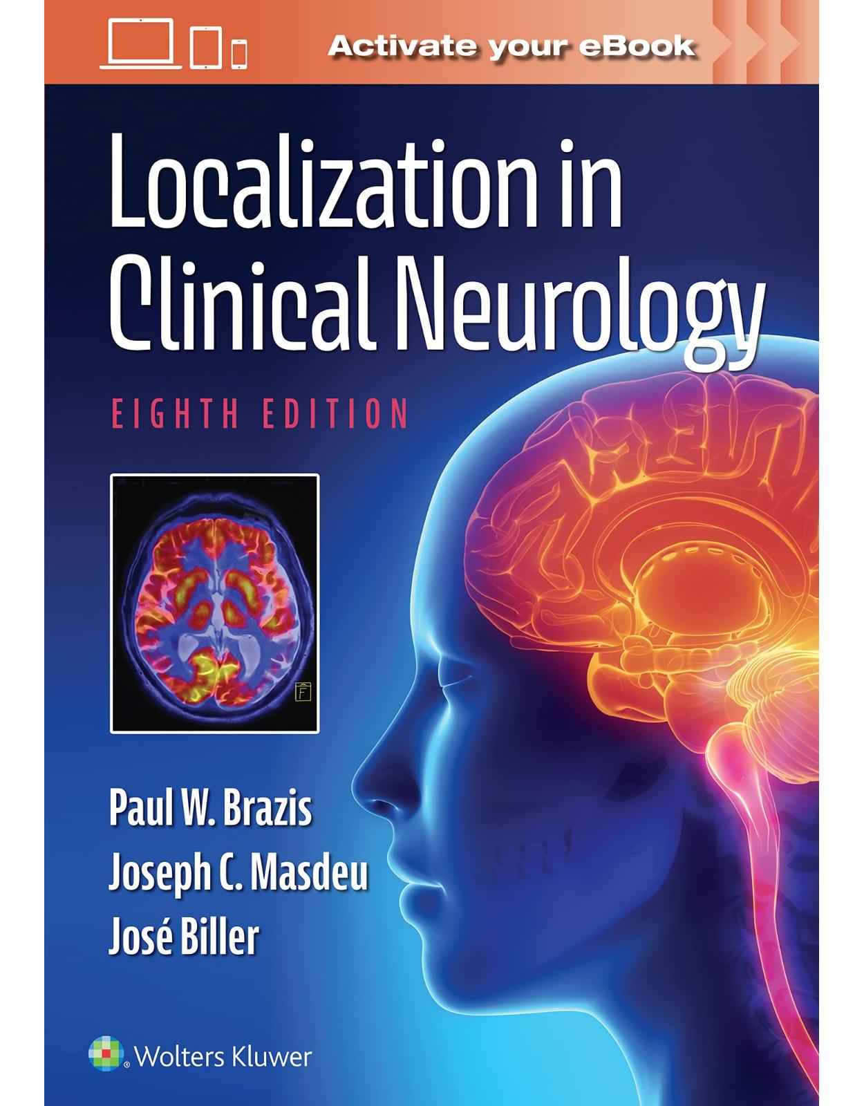
Localization in Clinical Neurology
Livrare gratis la comenzi peste 500 RON. Pentru celelalte comenzi livrarea este 20 RON.
Disponibilitate: La comanda in 3-4 saptamani
Editura: LWW
Limba: Engleza
Nr. pagini: 760
Coperta: Hardcover
Dimensiuni: 18.1 x 3.1 x 25.9 cm
An aparitie: 5 Oct. 2021
Description:
Anatomical localization skills based in physical examination are essential for any clinician caring for patients with neurologic disease processes. Now fully revised and up to date, Localization in Clinical Neurology, 8th Edition, uses easy-to-read descriptions, full-color illustrations and videos to help readers understand and locate the source of a patient’s signs and symptoms. This gold standard text now features dozens of clinical videos that help clinicians improve diagnostic accuracy and avoid unnecessary testing.
Enrich Your Digital Reading Experience
- Read directly on your preferred device(s), such as computer, tablet, or smartphone.
- Easily convert to audiobook, powering your content with natural language text-to-speech.
Table of Contents:
1 General Principles of Neurologic Localization
Introduction
A Brief History of Localization: Aphasia as an Example
Clinical Diagnosis and Lesion Localization
Localization of Lesions of the Motor System
Anatomy of the Motor System
Motor Signs and Symptoms and Their Localization
The Localization of Sensory Abnormalities
Anatomy of the Sensory System
Sensory Signs and Symptoms and Their Localization
Localization of Postural and Gait Disorders
Neural Structures Controlling Posture and Gait
Examination of Gait and Balance
Classification of Gait Disorders
2 Peripheral Nerves
Principal Signs and Symptoms of Peripheral Nerve Disease
Sensory Disturbances
Motor Disturbances
Disturbances of Muscle Stretch Reflexes
Vasomotor, Sudomotor, and Trophic Disturbances
Mononeuropathy Multiplex
Polyneuropathy
Lesions of Individual Nerves
Dorsal Scapular Nerve (C4–C5)
Subclavian Nerve (C5–C6)
Long Thoracic Nerve (C5–C7)
Suprascapular Nerve (C5–C6)
Subscapular Nerves (C5–C7)
Thoracodorsal Nerve (C6–C8)
Anterior Thoracic Nerves (C5–T1)
Axillary Nerve (C5–C6)
Musculocutaneous Nerve (C5–C7)
Median Nerve (C6–T1)
Ulnar Nerve (C7–T1)
Nerve Lesions
Radial Nerve (C5–C8)
Medial Cutaneous Nerves of the Arm and Forearm (C8–T1)
Intercostobrachial Nerve (T2)
Iliohypogastric (T12–L1), Ilioinguinal (L1), and Genitofemoral (L1–L2) Nerves
Femoral Nerve (L2–L4)
Obturator Nerve (L2–L4)
Lateral Femoral Cutaneous Nerve (L2–L3)
Gluteal Nerves (L4–S2)
Posterior Femoral Cutaneous Nerve (S1–S3)
Pudendal Nerve (S1–S4)
Sciatic Nerve (L4–S3) and Its Branches
3 Cervical, Brachial, and Lumbosacral Plexuses
The Cervical Plexus
Anatomy
Lesions of the Cervical Plexus
The Brachial Plexus
Anatomy
Lesions of the Brachial Plexus
Neuralgic Amyotrophy
Total Plexus Paralysis
Upper Plexus Paralysis (Erb–Duchenne Type)
Middle Plexus Paralysis
Lower Plexus Paralysis (Déjerine-Klumpke Type)
Lesions of the Cords of the Brachial Plexus
Brachial Mononeuropathies
Thoracic Outlet Syndrome (Cervicobrachial Neurovascular Compression Syndrome)
The Lumbosacral Plexus
Anatomy
Lesions of the Lumbosacral Plexus
Lesions of the Entire Lumbosacral Plexus
Lesions of the Lumbar Segments
Lesions of the Sacral Plexus
4 Spinal Nerve and Root
Anatomy of the Spinal Nerves and Roots
Principles of Spinal Nerve and Root Localization
Sensory Symptoms
Motor Signs
Reflex Signs
Etiologies of Spinal Nerve and Root Lesions
The Localization of Nerve Root Syndromes
Lesions Affecting the Cervical Roots
Lesions Affecting the Thoracic Roots
Lesions of the Lumbar and Sacral Roots
The Localization of Lumbosacral Disc Disease
5 Spinal Cord
Anatomy of the Spinal Cord
Gross Anatomy and Relationship to Vertebral Levels
Cross-Sectional Anatomy of the Spinal Cord
Major Ascending and Descending Tracts of the Spinal Cord
Corticospinal Tract
Corticorubrospinal Tract
Lateral Reticulospinal Tract
Vestibulospinal Tract
Medial Reticulospinal Tract
Arterial Supply to the Spinal Cord
Venous Drainage of the Spinal Cord
Physiology of the Spinal Cord Circulation
Spinal Cord Lesions
Complete Spinal Cord Transection (Transverse Myelopathy)
Lesions Affecting the Spinal Cord Centrally
Posterolateral Column Disease
Posterior Column Disease
Anterior Horn Cell Syndromes
Combined Anterior Horn Cell and Corticospinal Tract Disease
Vascular Disorders of the Spinal Cord and Spinal Canal
Arterial Spinal Cord Infarction
Venous Spinal Cord Infarction
Spinal Cord Vascular Malformations
Spinal Canal Hemorrhages
Extramedullary Cord Lesions and Their Differentiation from Intramedullary Cord Lesions
Localization of Spinal Cord Lesions at Different Levels
Foramen Magnum Syndrome and Lesions of the Upper Cervical Cord
Lesions of the Fifth and Sixth Cervical Segments
Lesions of the Seventh Cervical Segment
Lesions of the Eighth Cervical and First Thoracic Segments
Lesions of the Thoracic Segments
Lesions of the First Lumbar Segment
Lesions of the Second Lumbar Segment
Lesions of the Third Lumbar Segment
Lesions of the Fourth Lumbar Segment
Lesions of the Fifth Lumbar Segment
Lesions of the First and Second Sacral Segments
Conus Medullaris Lesions
Cauda Equina Lesions
Neurogenic Bladder with Spinal Cord Lesions
Sexual Function
Fecal Incontinence
6 Cranial Nerve I (The Olfactory Nerve)
Anatomy of the Olfactory Pathways
Localization of Lesions Affecting the Olfactory Nerve
Lesions Causing Anosmia
The Foster–Kennedy Syndrome
Lesions Causing Parosmia and Cacosmia
7 Visual Pathways
Anatomy of the Visual System
The Retina
The Optic Nerves and Optic Chiasm
The Optic Tracts and Lateral Geniculate Bodies
The Optic Radiations
The Visual Cortex and Visual Association Areas
Vascular Supply of the Visual Pathways
Localization of Lesions in the Optic Pathways
Changes in Visual Perception
Types of Visual Field Defects
Localization of Visual Field Defects
Objective Findings with Lesions of the Optic Pathways
Optic Neuropathy
8 The Localization of Lesions Affecting the Ocular Motor System
Ocular Motor Muscles and Nerves
Orbital Muscles
Diplopia
Testing for Diplopia
Childhood Strabismus
Disease of the Ocular Muscles
Retinal Disease Causing Diplopia
Ocular Motor Nerves and Localization of Lesions
The Pupil
Supranuclear Control of Eye Movements
The Vestibular System
Full-Field Optokinetic Reflex
Smooth Pursuit System
Anatomy of the Pursuit System
Lesions Affecting Smooth Pursuit
The Saccadic System
Mechanical Properties of Saccadic Eye Movements
Convergence System
Fixation System
Gaze Palsies
Nystagmus and Other Ocular Oscillations
Oscillopsia
Optokinetic Drum
Jerk Nystagmus
Systems Classification of Nystagmus
Vestibular Nystagmus
Gaze-Holding Nystagmus
Visual Stabilization Nystagmus
Clinical Classification of Nystagmus
Saccadic Intrusions
Lid Nystagmus
The Eyelids
Ptosis
Eyelid Retraction and Lid Lag
9 Cranial Nerve V (The Trigeminal Nerve)
Anatomy of Cranial Nerve V (Trigeminal Nerve)
Motor Portion
Sensory Portion
Clinical Evaluation of Cranial Nerve V Function
Sensory Evaluation
Motor Evaluation
Reflex Evaluation
Localization of Lesions Affecting Cranial Nerve V
Supranuclear Lesions
Nuclear Lesions
Lesions Affecting the Preganglionic Trigeminal Nerve Roots
Lesions Affecting the Gasserian Ganglion
Raeder Paratrigeminal Syndrome
Gradenigo Syndrome
The Cavernous Sinus Syndrome
The Superior Orbital Fissure Syndrome
Lesions Affecting the Peripheral Branches of the Trigeminal Nerve
Jaw Drop
10 Cranial Nerve VII (The Facial Nerve)
Anatomy of Cranial Nerve VII (Facial Nerve)
Motor Division
Nervus Intermedius
Anatomy of the Peripheral Course of the Facial Nerve
Vascular Supply of the Facial Nerve
Clinical Evaluation of CN VII Function
Motor Function
Sensory Function
Reflex Function
Parasympathetic Function
Localization of Lesions Affecting CN VII
Supranuclear Lesions (Central or Upper Motor Neuron Facial Paralysis)
Nuclear and Fascicular Lesions (Pontine Lesions)
Posterior Fossa Lesions (Cerebellopontine Angle Lesions)
Lesions Affecting the Meatal Segment of the Facial Nerve in the Temporal Bone
Lesions Affecting the Facial Nerve within the Facial Canal Distal to the Meatal Segment but Proximal to the Exit of the Nerve to the Stapedius Muscle
Lesions Affecting the Facial Nerve within the Facial Canal Between the Exit of the Nerve to the Stapedius and the Exit of the Chorda Tympani
Lesions Affecting the Facial Nerve in the Facial Canal Distal to the Exit of the Chorda Tympani
Lesions Distal to the Stylomastoid Foramen
Abnormalities of Tear Secretion
Abnormalities of Eyelid Closure
Eyelid Closure Insufficiency
Excessive Eyelid Closure and Blepharospasm
Abnormal Facial Movements and Their Localization
Dyskinetic Movements
Dystonic Movements (Blepharospasm and Blepharospasm with Oromandibular Dystonia)
Hemifacial Spasm
Postparalytic Spasm and Synkinetic Movements
Miscellaneous Movements
11 Cranial Nerve VIII (The Vestibulocochlear Nerve)
Anatomy of Cranial Nerve VIII
Auditory Pathways
The Vestibular System
Clinical Evaluation of Cranial Nerve VIII Function
Sensorineural Deafness
Vertigo and Vestibular Function
Localization of Lesions Causing Deafness and Vertigo
Localization of Lesions Causing Sensorineural Deafness
Localization of Lesions Causing Vertigo
Benign Paroxysmal Positioning Vertigo
Peripheral Vestibulopathy
Ménière Disease
Vertigo Secondary to Middle Ear Disease
Vertigo Secondary to Viral Infections
Vertigo Secondary to Trauma
Vascular Causes of the Central Vestibular Syndrome
Multiple Sclerosis
Wernicke Encephalopathy
Cerebellopontine Angle Tumors
Vestibular Epilepsy
Other Central Nervous System Disorders
Systemic Causes of Dizziness and Vertigo
12 Cranial Nerves IX and X (The Glossopharyngeal and Vagus Nerves)
Anatomy of Cranial Nerve IX (Glossopharyngeal Nerve)
Clinical Evaluation of Cranial Nerve IX
Motor Function
Sensory Function
Reflex Function
Autonomic Function
Localization of Lesions Affecting the Glossopharyngeal Nerve
Supranuclear Lesions
Nuclear and Intramedullary Lesions
Extramedullary Lesions
Glossopharyngeal (Vagoglossopharyngeal) Neuralgia
Anatomy of Cranial Nerve X (Vagus Nerve)
Clinical Evaluation of Cranial Nerve X
Motor Function
Sensory Function
Reflex Function
Localization of Lesions Affecting the Vagus Nerve
Supranuclear Lesions
Nuclear Lesions and Lesions within the Brainstem
Lesions within the Posterior Fossa
Lesions Affecting the Vagus Nerve Proper
Lesions of the Superior Laryngeal Nerve
Lesions of the Recurrent Laryngeal Nerve
Syncope from Glossopharyngeal or Vagal Metastasis
Arnold Nerve Cough Reflex
13 Cranial Nerve XI (The Spinal Accessory Nerve)
Anatomy of Cranial Nerve XI (Spinal Accessory Nerve)
Clinical Evaluation of Cranial Nerve XI Function
Sternocleidomastoid Muscle
Trapezius Muscle
Localization of Lesions Affecting Cranial Nerve XI
Supranuclear Lesions
Nuclear Lesions
Infranuclear Lesions
14 Cranial Nerve XII (The Hypoglossal Nerve)
Anatomy of Cranial Nerve XII (The Hypoglossal Nerve)
Clinical Evaluation of Cranial Nerve XII
Localization of Lesions Affecting Cranial Nerve XII
Supranuclear Lesions
Nuclear Lesions and Intramedullary Cranial Nerve XII Lesions
Peripheral Lesions of Cranial Nerve XII
Abnormal Tongue Movements
Dysarthria
15 Brainstem
Medulla Oblongata
Anatomy of the Medulla
Vascular Supply of the Medulla
Medullary Syndromes
The Pons
Anatomy of the Pons
Vascular Supply of the Pons
Pontine Syndromes
The Mesencephalon
Anatomy of the Mesencephalon
Vascular Supply of the Mesencephalon
Mesencephalic Syndromes
16 The Cerebellum
Anatomy of the Cerebellum
Vascular Supply of the Cerebellum
Clinical Manifestations of Cerebellar Dysfunction
Hypotonia
Cerebellar Ataxia
Cerebellar Dysarthria
Tremor
Ocular Motor Dysfunction
Cerebellar Fits
Miscellaneous
Nonmotor Manifestations
Cerebellar Syndromes
Rostral Vermis Syndrome
Caudal Vermis Syndrome
Hemispheric Syndrome
Pancerebellar Syndrome
Syndromes of Cerebellar Infarction
Inferior Cerebellar Infarction (Posterior Inferior Cerebellar Artery)
Ventral Cerebellar Infarction (Anterior Inferior Cerebellar Artery)
Dorsal Cerebellar Infarct (Superior Cerebellar Artery)
17 The Localization of Lesions Affecting the Hypothalamus and Pituitary Gland
Anatomy of the Region
Main Hypothalamic Nuclear Groups
Connections of the Hypothalamus
Clinical Manifestations of Hypothalamic or Pituitary Dysfunction
Disturbances of Temperature Regulation
Disturbances of Alertness and Sleep
Autonomic Disturbances
Disturbances of Water Balance
Disturbances of Caloric Balance and Feeding Behavior
Disturbances of Reproductive Functions
Other Endocrine Disturbances
Disturbances of Memory
Disturbances of Emotional Behavior and Affect
Gelastic Seizures
Headache
Chronic Pain
Impaired Visual Acuity, Visual Field Defects
Diplopia, Pupillary Changes
Clinical Findings Resulting from Lesions in Various Areas of the Hypothalamus and in the Pituitary Gland
18 The Anatomic Localization of Lesions in the Thalamus
Functional Anatomy of the Thalamus
Vascular Supply of the Thalamus
Localization of Ischemic Thalamic Lesions
Paramedian Territory
Thalamogeniculate (Lateral Thalamic or Inferolateral Thalamic) Territory
Tuberothalamic (Anterolateral Thalamic) Territory
Territory of the Posterior Choroidal Arteries
Clinical Manifestations of Lesions in the Thalamus
Disturbances of Alertness
Autonomic Disturbances
Disturbances of Mood and Affect
Memory Disturbances
Sensory Disturbances
Motor Disturbances
Disturbances of Ocular Motility
Disturbances of Complex Sensorimotor Functions
Disturbances of Executive Function
Topographic Localization of Thalamic Lesions
Anterior Thalamic Region
Medial Thalamic Region
VL Thalamic Region
Posterior Region
19 Basal Ganglia
Anatomy of the Basal Ganglia
Inputs into the Striatum (Caudate and Putamen)
Striatal Efferents
Pallidal Afferents and Efferents
Nigral Afferents and Efferents
Lesions of the Basal Ganglia
Dyskinesias
Chorea
Tardive Dyskinesia and Other Tardive Syndromes
Orofacial Dyskinesia
Abdominal Dyskinesias
Ballism
Akathisia
Athetosis
Dystonia
Torticollis
Paroxysmal Dyskinesias
Myoclonus
Painful Legs and Moving Toes
Restless Legs Syndrome and Periodic Limb Movements of Sleep
Tics
Tremor
Hypokinetic and Bradykinetic Disorders
Parkinsonism
Stiff-Man (Stiff-Person) Syndrome
Cortical-Basal Ganglionic (Corticobasal) Degeneration
Progressive Supranuclear Palsy (Steele–Richardson–Olszewski Syndrome)
Dementia with Lewy Bodies
Multiple System Atrophy
Paraneoplastic Movement Disorders
20 The Localization of Lesions Affecting the Cerebral Cortex
Anatomy of the Cerebral Cortex
Symptoms and Signs Caused by Cerebral Cortical Lesions
Vegetative Disturbances
Disturbances of Attention
Emotional Disturbances
Memory Disturbances
Sensory Disturbances
Disturbances of Sensorimotor Integration and of Movement Execution (Parietal, Frontal)
21 Localization of Lesions in the Autonomic Nervous System
Organization of the Autonomic Nervous System
Sympathetic Nervous System
Parasympathetic Nervous System
Enteric Nervous System
22 Vascular Syndromes of the Forebrain, Brainstem, and Cerebellum
Arterial Blood Supply
The Internal Carotid Artery
Anterior Cerebral Artery
Middle Cerebral Artery
Posterior Cerebral Artery
Collateral Circulation
Syndromes of the Cerebral Arteries
Transient Ischemic Attacks
The Carotid Artery Syndrome
The AChA Syndrome
ACA Syndrome
MCA Syndrome
Lacunar Infarctions
Cerebral Hemorrhage Syndromes
General Features
Specific Signs by Location
Syndromes Related to Cerebral Aneurysms
Cavernous Internal Carotid Artery Aneurysms
Posterior Communicating Artery Aneurysms
MCA Aneurysms
Vertebrobasilar Circulation Aneurysms
Subarachnoid Hemorrhage
23 The Localization of Lesions Causing Coma
The Unresponsive Patient
Anatomic Substrate of Alertness
Signs with Localizing Value in Coma
Respiratory Patterns
Temperature Changes
The Pupils
Eye Movements
Corneal Reflex
Motor Activity of the Body and Limbs
Clinical Presentations of Coma-Inducing Lesions Depending on Their Location
Supratentorial Structural Lesions
Subtentorial Structural Lesions
Psychogenic Unresponsiveness
Diagnosis of Death by Neurologic Criteria
Index
| An aparitie | 5 Oct. 2021 |
| Autor | Paul W. Brazis, Joseph C. Masdeu MD, PhD, Jose Biller |
| Dimensiuni | 18.1 x 3.1 x 25.9 cm |
| Editura | LWW |
| Format | Hardcover |
| ISBN | 9781975160241 |
| Limba | Engleza |
| Nr pag | 760 |
| Versiune digitala | DA |


Clientii ebookshop.ro nu au adaugat inca opinii pentru acest produs. Fii primul care adauga o parere, folosind formularul de mai jos.