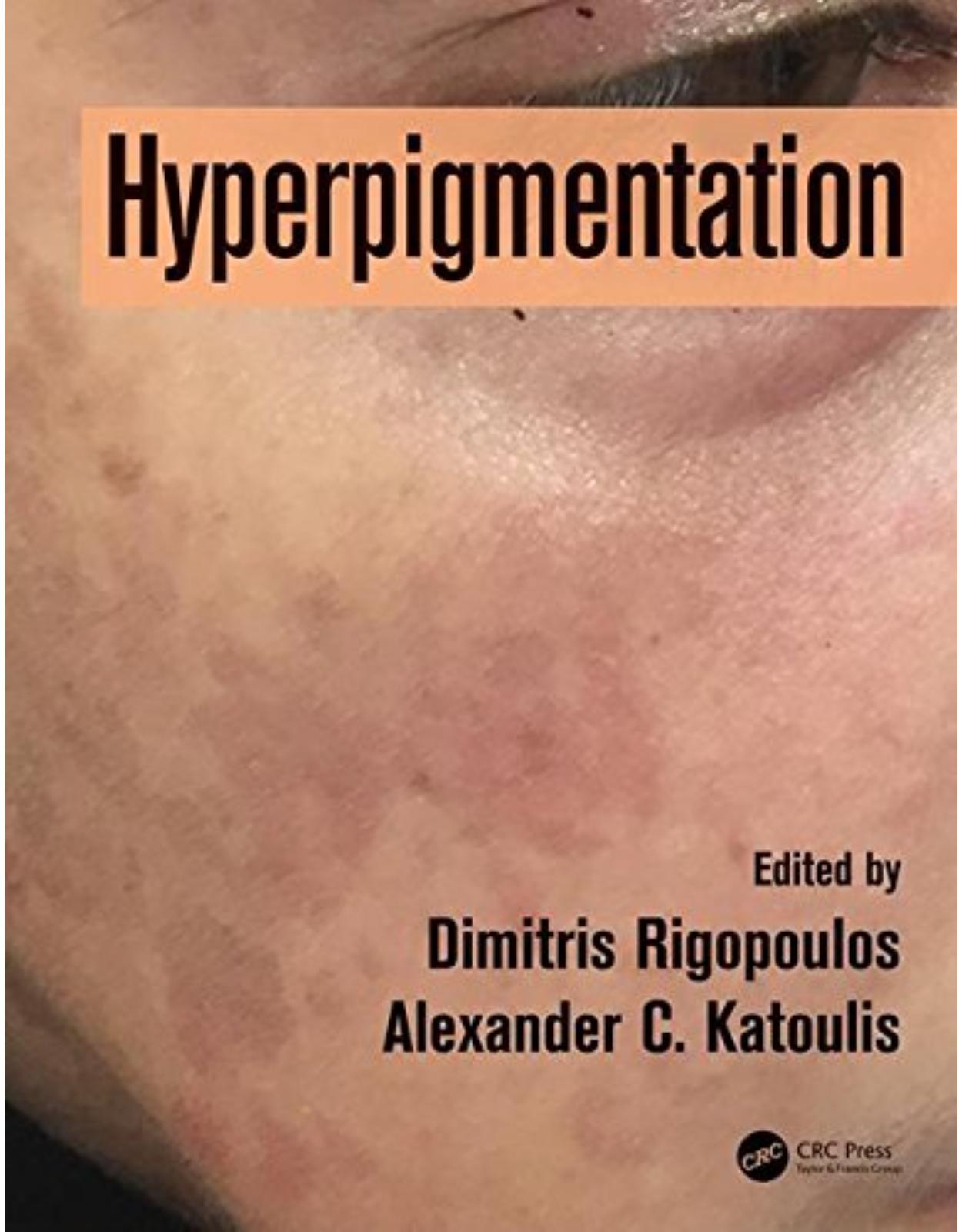
Hyperpigmentation
Livrare gratis la comenzi peste 500 RON. Pentru celelalte comenzi livrarea este 20 RON.
Disponibilitate: La comanda in aproximativ 4-6 saptamani
Editura: CRC Press
Limba: Engleza
Nr. pagini: 302
Coperta: Hardback
Dimensiuni: 1.3 x 15.9 x 22.9 cm
An aparitie: 17 October 2017
Description:
Darkening patches on the skin can result in great distress for the patient. However, definitive diagnosis between the many possible causes and effective treatment can be very hard for the medical practitioner to achieve. This is the first major professional reference with systematic coverage of diagnosis and the various treatment options, and it will be an indispensable clinical reference.
Table of Contents:
Section I Histology, Physiology, and Pathophysiology of Skin Pigmentation
Chapter 1 Color of skin
Introduction
Biology of skin color
Mechanism of melanin synthesis
Melanosome transport and transfer to keratinocytes
Racial and ethnic differences in skin color
Geographic variation
Genetics of skin color
Sexual dysmorphism
Skin color and skin typing
Measurement of skin color
Skin color and vitamin D
Skin color and folate
Skin color and photoaging
Skin color and cancer
Skin color and tanning
Skin color and discrimination
Disorders of skin color
References
Chapter 2 The melanocyte and melaninogenesis
The melanocyte
Melaninogenesis
Pathways regulating melaninogenesis
Regulatory proteins
Ultraviolet radiation
Paracrine melanogenic regulators
Other melanogenic stimulators
Melanogenic inhibitors
References
Chapter 3 Melanin
Historical background
Melanin
Chemical and physical properties
Site of melanin synthesis and storage
Extracutaneous melanin
Melanin metabolism
Detection of melanin (Special Stains for melanin)
Melanin pigment functions in health and diseases
Normal variations and differences in skin melanization
Gender
Age
Health
Melanin role in pigmentary and nonpigmentary disorders
References
Chapter 4 Pathophysiology of hyperpigmentation
References
SectionIIDisorders of Hyperpigmentation
Chapter 5 Melasma
Introduction
Pathogenesis
Clinical presentation
Diagnosis and differential diagnosis
Treatments for melasma
General measures and sun protection
Topical bleaching agents
Hydroquinone
Nonphenolic compounds
Combination formulas
Lasers and other therapies
Experimental therapies (
Maintenance treatment for melasma
References
Chapter 6 Erythema dyschromicum perstans
Introduction
History
Epidemiology
Pathophysiology
Clinical presentation
Histology
Laboratory findings
Differential diagnosis
Treatment
References
Chapter 7 Lichen planus pigmentosus
Introduction
Epidemiology
Etiology: Pathogenesis
Clinical manifestation
Histopathology
Dermatoscopy
Diagnosis
Differential diagnosis
Therapy
Prognosis
References
Chapter 8 Primary localized cutaneous amyloidosis
Introduction
Etiology: Pathogenesis
Clinical manifestations
Histopathology
Macular and lichen amyloidosis
Nodular amyloidosis
Use of special stains
Treatment
References
Chapter 9 Mastocytosis
Terminology and classification of mastocytosis
Pathogenesis
Molecular mechanisms
Heritability
Clinical manifestations
Skin signs and symptoms
Maculopapular cutaneous mastocytosis
Diffuse cutaneous mastocytosis
Skin mastocytomas
Symptoms rising from mediator release
Allergic reactions and anaphylaxis
Gastrointestinal symptoms
Neuropsychiatric symptoms
Musculosceletal symptoms
Therapeutical interventions in cutaneous mastocytosis
General measures
Pharmacological treatment
Prognosis
Infants and young children of
Older children and adults
References
Chapter 10 Pityriasis versicolor
Introduction
Microbiology (
Pathogenesis
Clinical features
Diagnosis
Confirmation of the diagnosis
Role of culture
Differential diagnosis
Treatment
Topical treatments
Nonspecific antifungal agents
Specific topical antifungal agents
Oral treatments
Prophylactic treatment
References
Chapter 11 Atrophoderma of Pasini Pierini
Introduction
Causes: Pathogenesis
Physical
Histological features
Laboratory studies
Differential diagnosis
Comorbidities
Treatment
Follow-up
References
Chapter 12 Poikiloderma of Civatte
Introduction
Epidemiology
Etiology and pathogenesis
Clinical presentation and course
Histopathology and electron microscopy
Differential diagnosis
Treatment
Prevention
References
Chapter 13 Pigmented nonmelanocytic skin lesions
Introduction
Lentigo
Lentigo simplex
Solar lentigo
Ink-spot lentigo
Oral and labial melanotic macules
Vulvar and penile lentigo
Syndromes characterized by the development of multiple lentigines
Xeroderma pigmentosum
LEOPARD syndrome
Peutz–Jeghers syndrome
Laugier–Hunziker syndrome
Myxoma syndrome
Carney syndrome
Dermoscopy features
Seborrheic keratosis
Clinical features
Histopathology
Dermoscopy features
Acanthotic type
Reticulated type
Verrucous type
Clonal SK
Melanoacanthoma
Lichen planus–like keratosis
Irritated SK
Pigmented basal cell carcinoma
Nevoid basal cell carcinoma syndrome
Bazex syndrome
Histopathology
Clinical presentation
Dermoscopy
Pigmented actinic keratosis
Clinical presentation
Histopathology
Dermoscopy
Pigmented squamous cell carcinoma
Clinical presentation
Histopathology
Dermoscopy
Pigmented intraepidermal carcinoma
Dermoscopy of invasive squamous cell carcinoma
Dermatofibroma
Clinical presentation
Histopathology
Dermoscopy
References
Chapter 14 Drug-induced hyperpigmention
Introduction
Pathogenesis
Clinical manifestation and course
Main drugs implicated in causing skin pigmentation
Tetracycline and tetracycline group
Retrovirals
NSAIDs
Antimalarials
Antiarrhythmics
Cytotoxic drugs
Metals and heavy metals
Psychotropic drugs
Anticonvulsants
ACTH
Carotene
Diagnostic points
Morphology
Treatment (
References
Chapter 15 Postinflammatory hyperpigmentation
Background
Epidemiology
Etiology
Pathogenesis
Clinical features
Diagnosis
Differential diagnoses
Therapy
Conclusion
References
Chapter 16 Linear hyperpigmentation
Pigmentary demarcation lines (futcher’s line and voigt’s line)
Definition
Epidemiology
Pathophysiology
Clinical features
Classification and location
Extracutaneous signs
Histopathology
Differential diagnoses
Treatment
Linear and whorled nevoid hypermelanosis
Definition
Epidemiology
Pathophysiology
Clinical features
Extracutaneous signs
Histopathology
Differential diagnoses
Treatment
Linear morphea or en coup de sabre
Definition
Epidemiology
Pathophysiology
Clinical features
Extracutaneous signs
Histopathology
Differential diagnoses
Treatment
Acquired Blaschkoid dermatitis
Definition
Epidemiology
Pathophysiology
Clinical features
Extracutaneous signs
Histopathology
Differential diagnoses
Treatment
Linear epidermal nevus
Definition
Epidemiology
Pathophysiology
Clinical features
Epidermal nevus syndromes
Complications of epidermal nevi
Extracutaneous signs
Histopathology
Differential diagnoses
Treatment
Becker’s nevus
Definition
Epidemiology
Pathophysiology
Clinical features
Malignancy
Extracutaneous signs
Histopathology,
Differential diagnoses
Treatment
Linear lichen planus
Definition
Epidemiology
Pathophysiology
Clinical features
Extracutaneous signs
Histopathology
Differential diagnoses
Treatment
Linea nigra
Definition
Epidemiology
Pathophysiology
Clinical features
Extracutaneous signs
Histopathology
Differential diagnoses
Treatment
Linea fusca
Definition
Epidemiology
Pathophysiology
Clinical features
Extracutaneous signs
Histopathology
Differential diagnoses
Treatment
Lichen striatus
Definition
Epidemiology
Pathophysiology
Clinical features
Extracutaneous signs
Histopathology
Differential diagnoses
Treatment
Linear postinflammatory hyperpigmentation
Definition
Epidemiology
Pathophysiology
Clinical features
Extracutaneous signs
Histopathology
Differential diagnoses
Treatment
Linear hyperpigmentation following chemotherapy
Epidemiology
Pathophysiology
Clinical features
Extracutaneous signs
Histopathology
Differential diagnoses
Treatment
Pigmentary lines of the newborn
Definition
Epidemiology
Pathophysiology
Clinical features
Extracutaneous signs
Histopathology
Differential diagnoses
Treatment
Heel-line or sock-line hyperpigmentation
Definition
Epidemiology
Pathophysiology
Clinical features
Extracutaneous signs
Histopathology
Differential diagnoses
Treatment
References
Chapter 17 Flagellate hyperpigmentation
Introduction
Bleomycin-induced flagellate dermatitis
Flagellate dermatitis associated with dermatomyositis
Shiitake dermatitis (toxicoderma): flagellate dermatitis triggered by shiitake mushroom ingestion
Other etiologies
Treatment
References
Chapter 18 Reticular hyperpigmentation
Introduction
Histopathogy
Diseases presenting with reticular hyperpigmentation in childhood
Diseases presenting with localized distribution of reticulate hyperpigmentation
Reticulate acropigmentation of Dohi (OMIM: 127400)
X-linked reticulate pigmentary disorder (OMIM: 301220)
Incontinentia pigmenti (OMIM: 308300)
Diseases presenting with widespread distribution of reticulate hyperpigmentation
Dyschromatosis universalis hereditaria
Epidermolysis bullosa simplex with mottled pigmentation (OMIM: 131960)
Dermatopathia pigmentosa reticularis (OMIM: 125595)
Naegeli–Franceschetti–Jadassohn syndrome (OMIM: 161000)
Dyskeratosis congenita
Diseases presenting with reticular hyperpigmentation in adolescence or adulthood
Diseases presenting with localized distribution of reticulate hyperpigmentation
Acropigmentation of Kitamura (OMIM: 615537)
Dowling–Degos disease (OMIM: 179850)
Galli–Galli disease
Lichen planus pigmentosus
Confluent and reticulate papillomatosis of Gougerot and Carteaud (OMIM: 167900)
Ashy dermatosis: Erythema dyschromicum perstans
Prurigo pigmentosa
References
Chapter 19 Genodermatoses
Introduction
Disorders with Increased Number of Epidermal Melanocytes
Disorders with increased number of dermal melanocytes
Disorders with Increased Melanin Levels
Disorders With Pigment Incontinence
References
SectionIIIDiagnosis and Differential Diagnosis
Chapter 20 Clinical examination
Introduction
Clinical Exam in Hyperpigmentations
Acquired hyperpigmentations
Melasma
Riehl melanosis: Toxic melanodermatitis
Poikiloderma of Civatte
Infraorbital dark circles
Acanthosis nigricans
Confluent and reticulated papillomatosis
Postinflammatory hyperpigmentation
Conclusion
References
Chapter 21 Wood’s lamp
Introduction
Mechanism of Action
Disorders of Pigmentation
Hypocromic and acromic disorders
Hypercromic disorders
Porphyrias
Skin Infections
Fungi
Bacteria
Other Uses
Conclusion
References
Chapter 22 Dermoscopy
Melanotic macules
Solar lentigo
Ink-spot lentigo
Labial melanotic macule
Mucosal melanotic macule
Cutaneous mastocytosis
Primary cutaneous amyloidosis
Regressing lichen ruber planus: ashy dermatosis
Melasma
Pigmentation within a scar
Tinea nigra
Onychomycosis
Nevoid lentigo of the nail unit
Laugier–hunziker syndrome
Subungual hemorrhage and subungual hematoma
Drug-induced nail pigmentation
Exogenous blue pigmentation
References
Chapter 23 Histopathology
Introduction
Hyperpigmentation in the cornified layer
Self-tanning products
Terra firma-forme dermatosis (dermatosis neglecta)
Tinea nigra
Pityriasis versicolor
Pigmented cornification above melanocytic neoplasms
Hyperpigmentation of the epidermis
Melanotic macule of the lip and benign melanosis of the vulva and penis
Ink-spot lentigo
Café-au-lait macule
Ephelides
Melasma
Becker’s nevus
Acanthosis nigricans
Confluent and reticulate papillomatosis (Gougerot–Carteaud)
Dowling–Degos disease
Galli–Galli disease
Solar lentigo
Seborrheic keratosis
Dermatosis papulosa nigra
Epidermal nevus
Late-stage morphea
Dermatofibroma
Mastocytosis
Notalgia paraesthetica
Pigmentation in the dermis or deeper tissues
Postinflammatory hyperpigmentation
Ashy dermatosis (erythema dyschromicum perstans)
Prurigo pigmentosa, late
Macular amyloidosis
Incontinentia pigmenti, late
Pigmented purpuric dermatoses
Hemochromatosis
Nevus of Ota, nevus of Ito, and Mongolian spot
Ochronosis (alkaptonuria)
Exogenous ochronosis
Minocycline hyperpigmentation
Argyria
Tattoo
References
Chapter 24 Reflectance confocal microscopy
Introduction
RCM findings in melasma
Conclusions
References
Chapter 25 Spectrophotometry
Introduction
Tristimulus reflectance colorimetry
Narrowband spectrophotometers
Tristimulus colorimetry versus narrowband spectrophotometers
Siascopy
Spectrophotometry and hyperpigmentary disorders
Conclusions
References
Chapter 26 Diagnostic algorithms
Introduction
Epidemiology
Clinical findings
Localized hyperpigmentation
Diffuse hyperpigmentation
History
Histopathology
Wood’s lamp examination
Dermoscopy
Conclusions
References
Chapter 27 Differential diagnoses of circumscribed and diffuse hyperpigmentation
Introduction
Taking steps toward diagnosis in disorders of color
Step 1: Demarcation
Step 2: Presence at birth
Step 3: Clinical morphology
Distribution
Color
Infiltration
Border or margin
Step 4: Dermoscopic features
Dermoscopic structures
Vascular patterns in dermoscopy
Step 5: History
Medical history
History of the lesion
Describing color
Features and clues for the diagnosis: Assessment of the hyperpigmented lesions or disorders and differential diagnoses
References
SectionIVTreatment
Chapter 28 Cosmetic camouflage for pigmentation issues
Introduction
Skin Color Issues
Cosmetics and Camouflaging
Facial Foundation Formulations
Oil-based foundations
Water-based foundations
Oil-free foundations
Anhydrous foundations
Facial Foundation Pigments and Specialty Additives
Facial Foundation Camouflaging Properties
Facial Foundation Application Varieties
Camouflage cosmetic application
Adverse Reactions to Camouflage Cosmetics
Camouflage Cosmetics for Skin of Color
Summary
References
Chapter 29 Sunscreens
UV Light
Sunscreens
Mechanism
Agents
Organic screens
Para-aminobenzoic acid and derivatives
Cinnamates
Salicylates
Benzophenones
Other organic screens
Inorganic screens
Other agents
Side Effects
Use of sunscreens after treatment for hyperpigmentation
Sunscreen after laser treatment
Sunscreen and depigmenting cream
Sunscreen after chemical peels
Sunscreen as monotherapy for hyperpigmentation
Sunscreen as prevention after treatment for melasma
References
Chapter 30 Medical therapy
The Principles of Medical Therapy
Introduction
The process of melanin formation
Principles of therapy
Medical Therapy
Sunscreens and sun protection
Sunscreen agents
Topical therapies
Phenolic compounds
Hydroquinone
Mequinol
Retinoids
Tretinoin
Adapalene
Tazarotene
Corticosteroids
Combination therapies and galenic formulations
Azelaic acid
Kojic acid
Ascorbic acid
Undecylenoyl phenylalanine
Arbutin
Other topical depigmenting agents
Chemical peels
Alpha hydroxy peels
Beta hydroxy peels
Conclusions
References
Chapter 31 Chemical peels
Introduction
Definition and Classification of Chemical Peels
Peeling Procedure
Peeling Agents
Alpha-hydroxy acid/GA peels
Beta-hydroxy acids
TCA peels
Phenol peels/peels for photoaging
Conclusions
References
Chapter 32 Cryotherapy
Introduction
Principles Of Cryobiology
Cryogen Selection
Equipment
Dewars
Cryosurgical units
Attachments for cryosurgical units
Other equipment
Cryosurgical Techniques
Preoperative Care
Common Hyperpigmented Skin Conditions Amenable to Cryosurgical Treatment
Benign
Seborrheic keratosis (SK)
Postoperative care
Lentigo simplex, ephelides
Postoperative care
Premalignant Lesions
Actinic keratosis
Postoperative care
Field cancerization
Postoperative care
Malignant Lesions
Where Not To Use Cryosurgery
Possible Complications
References
Chapter 33 Electrosurgery
Introduction
Terminology
Monopolar vs. bipolar
Monoterminal vs. biterminal
Frequencies
Preoperative Evaluation and Preparation
Electrocautery
Types of electrosurgeries (
Electrodesiccation and electrofulguration
Electrocoagulation
Electrosection
Safety Concerns
Cardiac pacemakers/ICDs
Postoperative Management
Complications
Dyschromia and scarring
Thermoelectric burn
Smoke plume
Fire
References
Chapter 34 Lasers
Introduction
Melasma
Photoaging
Nevus of ota
Post-inflammatory hyperpigmentation
Acknowledgments
References
Chapter 35 Intense pulsed light
What is intense pulsed light?
IPL as a therapy for hyperpigmentation
Melasma
Lentigines and ephelides
Poikiloderma of Civatte
Melanocytic lesions and others
Advantages and disadvantages of IPL
Clinical considerations before treatment with IPL
Adverse effects and safety of IPL
References
Index
| An aparitie | 17 October 2017 |
| Autor | Dimitris Rigopoulos, Alexander C. Katoulis |
| Dimensiuni | 1.3 x 15.9 x 22.9 cm |
| Editura | CRC Press |
| Format | Hardback |
| ISBN | 9781498740173 |
| Limba | Engleza |
| Nr pag | 302 |


Clientii ebookshop.ro nu au adaugat inca opinii pentru acest produs. Fii primul care adauga o parere, folosind formularul de mai jos.