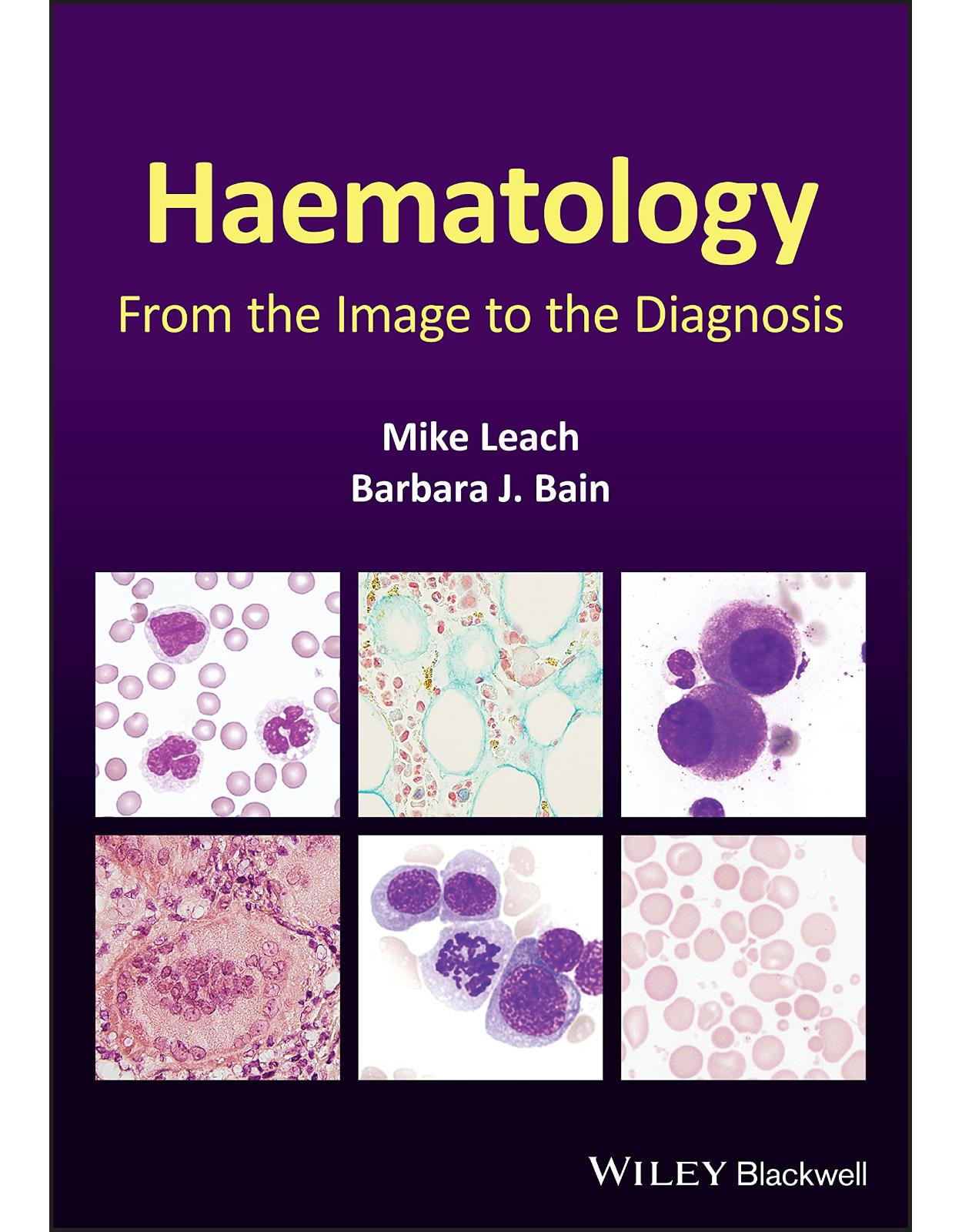
Haematology: From the Image to the Diagnosis
Livrare gratis la comenzi peste 500 RON. Pentru celelalte comenzi livrarea este 20 RON.
Disponibilitate: La comanda in aproximativ 4 saptamani
Autor: Mike Leach, Barbara J. Bain
Editura: Wiley
Limba: Engleza
Nr. pagini: 304
Coperta: Hardcover
Dimensiuni: 18.29 x 1.78 x 25.65 cm
An aparitie: 2 Sept. 2021
DESCRIPTION:
Haematology
Diagnostic haematology requires the assessment of clinical and laboratory data together with a careful morphological assessment of cells in blood, bone marrow and tissue fluids. Subsequent investigations including flow cytometry, immunohistochemistry, cytogenetics and molecular studies are guided by the original morphological findings. These targeted investigations help generate a prompt unifying diagnosis. Haematology: From the Image to the Diagnosis presents a series of cases illustrating how skills in morphology can guide the investigative process. In this book, the authors capture a series of images to illustrate key features to recognize when undertaking a morphological review and show how they can be integrated with supplementary information to reach a final diagnosis.
Using a novel format of visual case studies, this text mimics ‘real life’ for the practising diagnostic haematologist – using brief clinical details and initial microscopic morphological triage to formulate a differential diagnosis and a plan for efficient and economical confirmatory investigation to deduce the correct final diagnosis. The carefully selected, high-quality photomicrographs and the clear, succinct descriptions of key features, investigations and results will help haematologists, clinical scientists, haematology trainees and haematopathologists to make accurate diagnoses in their day-to-day work.
Covering a wide range of topics, and including paediatric as well as adult cases, Haematology: From the Image to the Diagnosis is a succinct visual guide which will be welcomed by consultants, trainees and scientists alike.
TABLE OF CONTENTS:
Preface
Abbreviations
1. Haemophagocytic syndrome secondary to anaplastic large cell lymphoma
2. Bone marrow AL amyloidosis
3. Cup-like blast morphology in acute myeloid leukaemia
4. Neutrophil morphology
5. Primary myelofibrosis
6. Sarcoidosis
7. Leishmaniasis
8. Gelatinous transformation of the bone marrow
9. Acanthocytic red cell disorders
10. Large granular lymphocytic leukaemia
11. Pure erythroid leukaemia
12. Reactive mesothelial cells
13. Plasmablastic myeloma
14. Septicaemia
15. Unstable haemoglobin (haemoglobin Köln) and a myeloproliferative neoplasm
16. Sickle cell anaemia in crisis
17. Acute myeloid leukaemia with t(8;21)(q22;q22.1)
18. Chronic neutrophilic leukaemia
19. Essential thrombocythaemia
20. Hairy cell leukaemia
21. Mantle cell lymphoma in leukaemic phase
22. Infantile osteopetrosis
23. Reactive eosinophilia
24. Stomatocytic red cell disorders
25. Reactive lymphocytosis due to viral infection
26. Therapy-related acute myeloid leukaemia with eosinophilia
27. Red cell fragmentation syndromes
28. NK/T-cell lymphoma in leukaemic phase
29. Myelodysplastic syndrome with del(5q)
30. Classical Hodgkin lymphoma
31. Cryoglobulinaemia
32. Congenital dyserythropoietic anaemia
33. Acute monoblastic leukaemia with t(9;11)(p21.3;q23.3)
34. Chronic myeloid leukaemia presenting with myeloid sarcoma and extreme thrombocytosis
35. Glucose-6-phosphate dehydrogenase deficiency
36. Leukaemic presentation of hepatosplenic gamma-delta T-cell lymphoma
37. Myelodysplastic syndromes
38. Pelger–Huët anomaly
39. Russell bodies in lymphoplasmacytic lymphoma
40. T-cell prolymphocytic leukaemia
41. Myeloid maturation arrest
42. MDS/MPN with ring sideroblasts and thrombocytosis
43. Acute myeloid leukaemia with inv(16)(p13.1q22)
44. Babesiosis
45. Haemoglobin E disorders
46. Juvenile myelomonocytic leukaemia
47. Non-haemopoietic tumours
48. Richter transformation of chronic lymphocytic leukaemia
49. Sickle cell-haemoglobin C disease
50. T cell/histiocyte-rich B-cell lymphoma
51. Miliary tuberculosis
52. Pure red cell aplasia
53. Lymphoblastic transformation of follicular lymphoma
54. Primary hyperparathyroidism
55. Gamma heavy chain disease
56. Acute promyelocytic leukaemia with t(15;17)(q24.1;q21.2)
57. AA amyloidosis
58. Acquired sideroblastic anaemia
59. Diffuse large B-cell lymphoma
60. Hickman line infection
61. Monocytes and their precursors
62. Paroxysmal cold haemoglobinuria
63. Transient abnormal myelopoiesis
64. Systemic lupus erythematosus
65. Granular blast cells in acute lymphoblastic leukaemia
66. Chronic myelomonocytic leukaemia
67. Burkitt lymphoma/leukaemia
68. Gaucher’s disease
69. Myelodysplastic syndrome with haemophagocytosis
70. Primary oxalosis
71. Acute myeloid leukaemia with inv(3)(q21.3q26.2)
72. Autoimmune haemolytic anaemia
73. Chronic eosinophilic leukaemia due to FIP1L1-PDGFRA fusion gene
74. Leukaemic phase of follicular lymphoma
75. Megaloblastic anaemia
76. Reactive bone marrow and an abnormal PET scan
77. Acute megakaryoblastic leukaemia
78. Erythrophagocytosis and haemophagocytosis
79. Hyposplenism
80. Acquired haemoglobin H disease
81. Cystinosis
82. Familial platelet disorder with a predisposition to AML
83. Nodular lymphocyte predominant Hodgkin lymphoma
84. Acute monocytic leukaemia with NPM1 mutation
85. Adult T-cell leukaemia/lymphoma
86. Hereditary elliptocytosis and pyropoikilocytosis
87. Sézary syndrome
88. Spherocytic red cell disorders
89. Acute myeloid leukaemia and metastatic carcinoma
90. Chédiak-Higashi syndrome
91. Cortical T-lymphoblastic leukaemia/lymphoma
92. Trypanosomiasis
93. Acute myeloid leukaemia with myelodysplasia-related changes
94. Blastic plasmacytoid dendritic cell neoplasm
95. Inherited macrothrombocytopenias
96. Persistent polyclonal B-cell lymphocytosis
97. Acute myeloid leukaemia with t(6;9)(p23;q34.1)
98. B-cell prolymphocytic leukaemia
99. Various red cell enzyme disorders
100. Sea blue histiocytosis in multiple myeloma
101. Enteropathy-associated T-cell lymphoma
Answers to multiple choice questions and further reflections on the theme
| An aparitie | 2 Sept. 2021 |
| Autor | Mike Leach, Barbara J. Bain |
| Dimensiuni | 18.29 x 1.78 x 25.65 cm |
| Editura | Wiley |
| Format | Hardcover |
| ISBN | 9781119777502 |
| Limba | Engleza |
| Nr pag | 304 |

