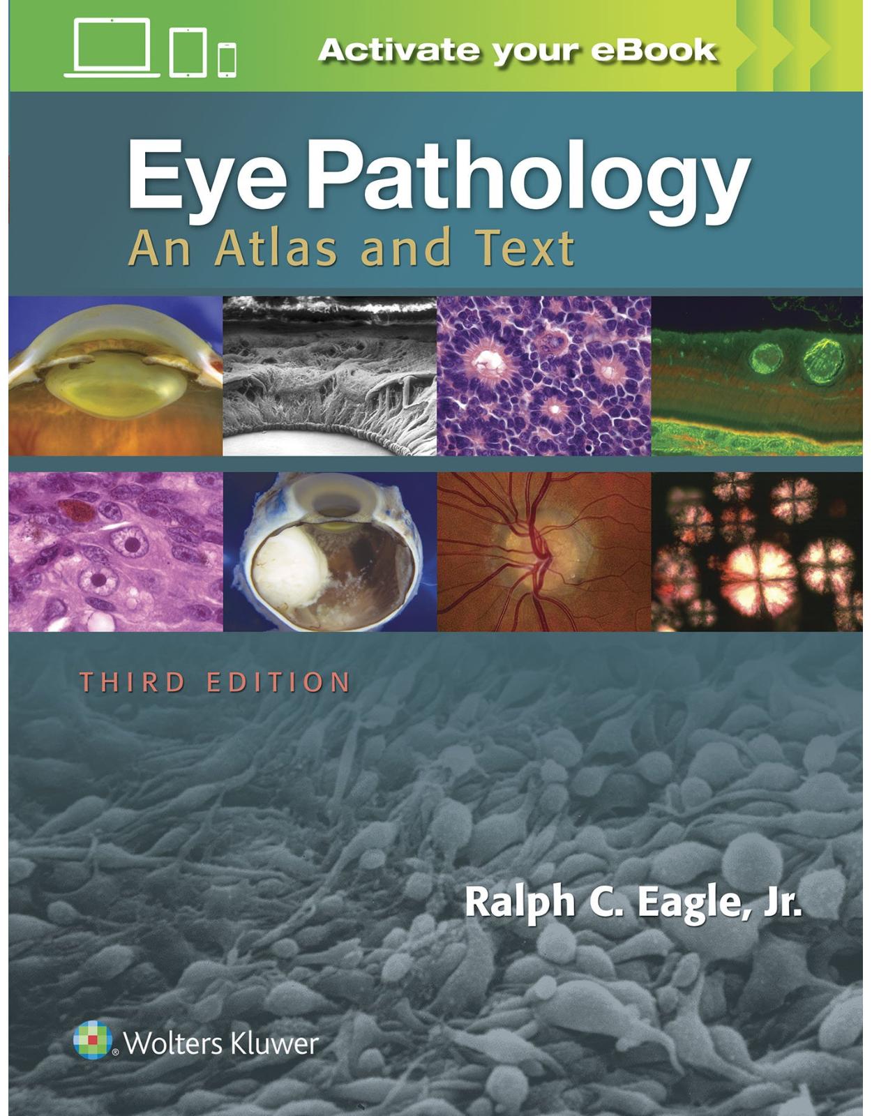
Eye Pathology: An Atlas and Text
Livrare gratis la comenzi peste 500 RON. Pentru celelalte comenzi livrarea este 20 RON.
Disponibilitate: La comanda in aproximativ 4-6 saptamani
Autor: Ralph C. Eagle
Editura: LWW
Limba: Engleza
Nr. pagini: 384
Coperta: Hardcover
Dimensiuni: 21.84 x 2.03 x 27.94 cm
An aparitie: 2016
Description:
Master the eye pathology you need to know for the OKAP exam, residency, and beyond! Here's a perfect introduction to basic eye pathology that can easily be read and mastered during an ophthalmic pathology rotation. It provides effective, efficient preparation for OKAP examinations or Board certification in ophthalmology, and will also serve as a concise clinical reference in practice. Richly illustrated and masterfully written, this best-selling ophthalmology resource equips you to understand eye pathology. Key Features:•Identify ocular diseases and disorders with the aid of more than 750 high-quality color illustrations.•Master the latest knowledge in the field with thorough updates on abusive head trauma; spectral domain optical coherence tomography (SD-OCT); the revised IC3D classification of corneal dystrophies; IgG4-related disease; the genetics of melanoma and retinoblastoma; interpretation of lamellar keratoplasty specimens from DALK, DSEK, and refractive surgical procedures; ;; immunohistochemistry; and much more.•Gauge your mastery of the material with more than 650 multiple-choice review questions online.Now with the print edition, enjoy the bundled interactive eBook edition, offering tablet, smartphone, or online access to: •Complete content with enhanced navigation•A powerful search that pulls results from content in the book, your notes, and even the web•Cross-linked pages, references, and more for easy navigation•Highlighting tool for easier reference of key content throughout the text•Ability to take and share notes with friends and colleagues•Quick reference tabbing to save your favorite content for future use
Table of Contents:
chapter01 An Introduction to Ocular Anatomy and Histology
chapter02 Congenital and Developmental Anomalies
chapter03 Inflammation
chapter04 Ocular Trauma
chapter05 Conjunctiva
chapter06 Cornea and Sclera
chapter07 The Lens
chapter08 Glaucoma
chapter09 Retina and Vitreous
chapter11 Intraocular Tumors in Adults
chapter12 Retinoblastoma and Simulating Lesions
chapter13 The Eyelid and Lacrimal Drainage System
chapter14 Orbit and Optic Nerve
chapter16 Laboratory Techniques, Special Stains, and Immunohistochemistry
Online Questions
Chapter 1: An Introduction to Ocular Anatomy and Histology
Chapter 1 Introduction
Figure 1-1
Figure 1-2: The iris.
Figure 1-3: The retina.
Figure 1-4: The retinal pigment epithelium (RPE).
Figure 1-5: Ciliary body.
Figure 1-6: The lens.
Figure 1-7: The suspensory ligament of the lens.
Figure 1-8: The cornea.
Figure 1-9: The iridocorneal angle and aqueous outflow pathways.
Figure 1-10: The optic nerve.
Bibliography
Chapter 2: Congenital and Developmental Anomalies
Chapter 2 Introduction
Figure 2-1: Trisomy 13.
Figure 2-2: Microphthalmos with colobomatous cyst.
Chromosomal Anomalies
Figure 2-3: Brushfield spots, trisomy 21.
Heritable Disorders with Ocular Manifestations
Figure 2-4: Aniridia.
Phakomatoses (Familial Tumor Syndromes)
Figure 2-5: Elephantiasis neuromatosa, von Recklinghausen neurofibromatosis (NF-1).
Figure 2-6: von Recklinghausen neurofibromatosis (NF-1) Choroidal infiltrate, NF-1.
Figure 2-7: Lisch nodules, NF-1.
Figure 2-8: Plexiform neurofibroma, NF-1.
Figure 2-9
Figure 2-10: Retinal hemangioblastoma, von Hippel-Lindau syndrome.
Figure 2-11: Sturge-Weber syndrome.
Figure 2-12: Cavernous hemangioma the retina.
Bibliography
General References
Chromosomal Anomalies
Aniridia and Other Iris Anomalies
Neurofibromatosis
Tuberous Sclerosis Complex
Von Hippel-Lindau Syndrome
Sturge-Weber Syndrome
Chapter 3: Inflammation
Introduction
Figure 3-1: Acute dacryocystitis.
The Classification of Inflammation
Polymorphonuclear Leukocytes
Figure 3-2: Inflammatory cells.
Eosinophils
Lymphocytes
Plasma Cells
Figure 3-3: Russell bodies.
Figure 3-4: Iritis.
Mast Cells
Figure 3-5
Macrophages
Figure 3-6: Macrophages.
Epithelioid Histiocytes and Giant Cells
Figure 3-7
Figure 3-8
Figure 3-9: Foreign body giant cell.
Granulomatous Inflammation
Patterns of Chronic Granulomatous Inflammation
Figure 3-10: Patterns of granulomatous inflammation.
Figure 3-11: Granulation tissue.
Endophthalmitis and Panophthalmitis
Figure 3-12: Vitreous abscess, acute bacterial endophthalmitis.
Figure 3-13: Fungal endophthalmitis.
Figure 3-14
Figure 3-15: Exogenous endophthalmitis due to infected filtering bleb.
Figure 3-16: Propionibacterium acnes localized endophthalmitis.
Figure 3-17: Endogenous fungal endophthalmitis, aspergillosis.
Figure 3-18: Endogenous endophthalmitis secondary to Nocardia.
Viral Retinitis
Figure 3-19: Cytomegalovirus retinitis.
Figure 3-20: Progressive Outer Retinal Necrosis (PORN) syndrome.
Toxoplasma Retinochoroiditis
Figure 3-21: Toxoplasma retinochoroiditis.
Uveitis
The Sequelae of Ocular Inflammation
Figure 3-22: Cyclitic membrane.
Figure 3-23
Bibliography
General References
Inflammatory Mediators
Inflammatory Cells
Granulomatous Inflammation
Bacterial Endophthalmitis
Fungal Endophthalmitis
HIV/AIDS
Viral Retinitis
Uveitis
Parasitic Infection
Chapter 4: Ocular Trauma
Chapter 4 Introduction
Figure 4-1
Figure 4-2
Figure 4-3
Figure 4-4: Spontaneous expulsive hemorrhage.
Sympathetic Uveitis (Sympathetic Ophthalmia)
Figure 4-5: Sympathetic uveitis.
Contusion Injuries
Figure 4-6: Anterior retinal avulsion, contusion injury.
Figure 4-7
Figure 4-8: Corneal blood staining.
Figure 4-9: Contusion injuries, anterior segment.
Intraocular Foreign Bodies
Figure 4-10: Intraocular foreign body (BB).
Figure 4-11: Siderosis.
Figure 4-12
Chemical Injuries
Figure 4-13: Alkali injury.
Wound Healing
Figure 4-14: Scar of corneal laceration.
Surgical Complications
Figure 4-15
Figure 4-16: Extramedullary hematopoiesis, traumatized eye with chronic intraocular hemorrhage.
Bibliography
Surgical Trauma
Nonsurgical Trauma
Sympathetic Uveitis
Intraocular Foreign Bodies
Chemical Burns
Radiation Injuries
Chapter 5: Conjunctiva
Chapter 5 Introduction
Figure 5-1: Conjunctival epithelium.
Developmental Lesions
Figure 5-2: Developmental lesions.
Figure 5-3: Osseous choristoma, conjunctiva.
Conjunctivitis
Figure 5-4: Acute purulent conjunctivitis.
Figure 5-5: Ligneous conjunctivitis.
Chronic Conjunctivitis
Figure 5-6
Figure 5-7: Trachoma, conjunctival smear.
Figure 5-8: Conjunctival papillae.
Figure 5-9: Vernal conjunctivitis.
Chronic Granulomatous Conjunctivitis
Figure 5-10: Sarcoidosis.
Figure 5-11: Cat scratch disease, conjunctiva.
Figure 5-12: Synthetic fiber granuloma.
Figure 5-13
Figure 5-14: Ocular cicatricial pemphigoid.
Figure 5-15
Degenerations
Figure 5-16: Pinguecula.
Figure 5-17: Amyloidosis, conjunctiva.
Conjunctival Cysts
Conjunctival Neoplasms
Squamous Epithelial Lesions
Figure 5-18: Conjunctival papilloma.
Figure 5-19: Actinic keratosis, conjunctiva.
Figure 5-20: Squamous cell lesions, conjunctiva.
Figure 5-21: Squamous cell neoplasms.
Figure 5-22
Figure 5-23
Figure 5-24: Hereditary benign intraepithelial dyskeratosis, conjunctiva.
Pigmented Lesions of the Conjunctiva
Figure 5-25
Figure 5-26: Constitutional melanosis.
Figure 5-27: Hormone-induced changes in conjunctival nevus.
Figure 5-28: Compound cystic nevus, conjunctiva.
Figure 5-29: Congenital ocular melanocytosis.
Primary Acquired Melanosis (Intraepithelial Melanocytic Proliferation)
Figure 5-30: Primary acquired melanosis.
Conjunctival Melanoma
Figure 5-31: Malignant melanoma arising from PAM with atypia.
Lymphoid Neoplasms
Figure 5-32: Conjunctival MALT lymphoma.
Figure 5-33: Benign reactive lymphoid hyperplasia.
Figure 5-34: Follicular lymphoma.
Caruncular Lesions
Figure 5-35: Caruncular lesions.
Figure 5-36: Oncocytoma, caruncle.
Bibliography
Developmental Lesions
Conjunctivitis
Granulomatous Conjunctivitis
Parasitic and Mycotic Infections
Ocular Cicatricial Pemphigoid
Amyloidosis
Conjunctival Tumors
Squamous Epithelial Lesions
Melanocytic Lesions
Lymphoid Lesions
Miscellaneous Lesions
Tumors of the Caruncle
Chapter 6: Cornea and Sclera
Corneal Histology
Developmental Anomalies
Figure 6-1: Posterior embryotoxon.
Figure 6-2: Peters anomaly.
Figure 6-3: Aniridia keratopathy.
Corneal Inflammation
Acute Keratitis and Corneal Ulceration
Figure 6-4: Acute keratitis.
Figure 6-5: Pseudomonas sclerokeratitis.
Figure 6-6: Infectious pseudocrystalline keratopathy.
Figure 6-7: Fungal keratitis.
Figure 6-8: Acute keratitis secondary to Candida after DSEK.
Viral Keratitis
Figure 6-9: Herpes simplex keratitis.
Interstitial Keratitis
Figure 6-10: Chronic luetic interstitial keratitis.
Parasitic Keratitis
Figure 6-11: Microsporidia keratitis.
Figure 6-12: Acanthamoeba keratitis.
Peripheral Corneal Ulcers
Figure 6-13: Mooren ulcer.
Pterygium
Figure 6-14: Pterygium.
Other Corneal Degenerations
Figure 6-15: Band keratopathy.
Figure 6-16: Chronic actinic keratopathy.
Figure 6-17: Corneal staphyloma.
Figure 6-18: Keratomalacia secondary to avitaminosis
Figure 6-19: Degenerative pannus, chronic corneal edema.
Keratoconus
Figure 6-20: Keratoconus.
Figure 6-21: Keratoconus, characteristic dehiscences in the Bowman membrane.
Corneal Dystrophies
Epithelial and Subepithelial Dystrophies
Epithelial–Stromal Dystrophies Caused by Mutations in TGFBI
Stromal Dystrophies
Figure 6-22: Epithelial dystrophies.
Figure 6-23: Gelatinous drop-like dystrophy.
Figure 6-24
Figure 6-25: Lattice corneal dystrophy, type I.
Figure 6-26: Granular corneal dystrophy.
Figure 6-27: Granular dystrophy type 2 recurrent in flap after LASIK.
Figure 6-28: Macular corneal dystrophy.
Figure 6-29: Schnyder corneal dystrophy.
Corneal Edema, Bullous Keratopathy, and the Endothelial Dystrophies
Figure 6-30: Corneal edema.
Figure 6-31: Fuchs dystrophy.
Figure 6-32: Fuchs dystrophy, flat preparations of DSEK specimens.
Figure 6-33: Pseudophakic bullous keratopathy.
Figure 6-34: Posterior polymorphous dystrophy.
Corneal Manifestations of Systemic Disease
Figure 6-35: Corneal rings.
Figure 6-36: Immunoglobulin deposits in protein dyscrasias.
Corneal Transplantation
Figure 6-37: Lamellar keratoplasty.
The Sclera
Figure 6-38: Rheumatoid scleritis.
Bibliography
Developmental Lesions
Bacterial Keratitis
Herpetic Keratitis
Epidemic Keratoconjunctivitis
Interstitial Keratitis
Acanthamoeba Keratitis
Other Types of Parasitic and Protozoal Keratitis
Peripheral Corneal Ulcerations
Corneal Degenerations
Vitamin A Deficiency
Keratoconus
Corneal Dystrophies
Epithelial and Bowman’s Membrane Dystrophies
Stromal Dystrophies
Endothelial Dystrophies
The Cornea in Systemic Disease
Sclera
Chapter 7: The Lens
Chapter 7 Introduction
Congenital Anomalies of the Lens
Figure 7-1
Figure 7-2: Developmental cataracts.
Cataract
Figure 7-3: Pseudophakia.
Figure 7-4: Common types of cataract.
Figure 7-5: Nuclear sclerosis.
Figure 7-6: Oxalate crystals, nuclear sclerosis.
Figure 7-7: Cortical cataract.
Figure 7-8: Morgagnian cataract.
Figure 7-9: Anterior subcapsular cataract.
Figure 7-10: Capsular fibrosis, postextracapsular cataract extraction (post-ECCE).
Figure 7-11: Bladder cells, posterior subcapsular cataract.
Figure 7-12: Elschnig pearls, post-ECCE.
Complicated Cataracts
Figure 7-13: Fuchs heterochromic cyclitis.
Sugar Cataracts: Diabetes Mellitus and Galactosemia
Cataract and Systemic Disease
Traumatic Cataracts
Figure 7-14: Soemmerring ring cataract, post-ECCE.
Toxic Cataract
Lens Capsular Abnormalities
Figure 7-15: Pseudoexfoliation of the lens capsule.
Figure 7-16: Pseudoexfoliation syndrome, iris pigment epithelium.
Figure 7-17: Polychromasia capsulare.
Zonular Fibers and Ectopia Lentis
Figure 7-18: Ectopia lentis.
Bibliography
Developmental Anomalies
Rubella Cataract
Cataract in Genetic Syndromes
Senile Cataract
Posterior Capsular Opacification
Posterior Subcapsular Cataract
Cataracts Caused by Ocular Disease (Complicated Cataracts)
Sugar Cataracts
Cataracts Associated with Systemic Disease
Traumatic Cataract
Toxic Cataract
True Exfoliation of the Lens Capsule
Pseudoexfoliation of the Lens Capsule
Heritable Lens Dislocation (Ectopia Lentis)
Chapter 8: Glaucoma
Chapter 8 Introduction
Figure 8-1: Normal perifoveal retina (A) compared to glaucomatous retinal atrophy (B).
Figure 8-2
Figure 8-3: Glaucomatous optic atrophy, scanning electron micrograph (SEM).
Figure 8-4: Iridocorneal angle.
Figure 8-5
Classification of the Glaucomas
Figure 8-6: Open and closed anterior chamber angles.
Developmental Glaucoma
Figure 8-7: Congenital glaucoma.
Primary Open-Angle Glaucoma
Primary Closed-Angle Glaucoma
Secondary Closed-Angle Glaucoma
Figure 8-8: Iris bombe.
Figure 8-9: Epithelial downgrowth.
Figure 8-10: Neovascular glaucoma.
Figure 8-11: Neovascular glaucoma.
Figure 8-12: Neovascular glaucoma.
Figure 8-13
Figure 8-14: ICE syndrome, Cogan-Reese variant.
Neovascular Glaucoma
The Iridocorneal Endothelial (ICE) Syndrome
Secondary Open-Angle Glaucoma
Figure 8-15: Phacolytic glaucoma.
Figure 8-16
Figure 8-17: Pigmentary glaucoma-iris transillumination.
Figure 8-18: Pigmentary glaucoma.
Figure 8-19: Ring melanoma.
Figure 8-20: Secondary closed-angle glaucoma in eyes with choroidal melanomas and total retinal detachments.
Figure 8-21
Ocular Tissue Changes in Glaucomatous Eyes
Bibliography
Genetics and Pathogenesis
Developmental Glaucoma
Primary Open Angle Glaucoma
Angle Closure Glaucoma
Neovascular Glaucoma
The Iridocorneal Endothelial Syndrome
Phacolytic Glaucoma
Pigmentary Glaucoma
Pseudoexfoliation Syndrome
Other Secondary Glaucomas
Tumors and Glaucoma
Chapter 9: Retina
Chapter 9 Introduction
Developmental Anomalies
Figure 9-1: Albinism.
Retinal Hemorrhages and Exudates
Figure 9-2: Retinal hemorrhages.
Figure 9-3: Abusive head trauma (shaken baby syndrome).
Figure 9-4: Retinal exudates.
Figure 9-5: Macular star figure.
Figure 9-6: Cotton-wool spots.
Angioid Streaks
Figure 9-7: Angioid streaks.
Retinal Artery and Arteriolar Occlusions
Figure 9-8
Figure 9-9: Retinal opacification.
Figure 9-10: Giant cell arteritis, temporal artery biopsy.
Retinal Venous Occlusion
Figure 9-11: Central retinal vein occlusion.
Hypertensive Retinopathy
Retinal Arteriosclerosis
Figure 9-12: Retinal arteriolar sclerosis.
Cystoid Macular Edema
Figure 9-13: Cystoid macular edema.
Macular Holes
Age-Related Maculopathy (AMD; Age-Related Macular Degeneration, ARMD)
Figure 9-14
Figure 9-15: Atrophic form of age-related macular degeneration.
Figure 9-16: Exudative form of age-related macular degeneration.
Figure 9-17: Drusen.
Figure 9-18: Ocular histoplasmosis syndrome.
The Retinal Pigment Epithelium
Stargardt Disease (Fundus Flavimaculatus)
Figure 9-19: Stargardt disease (fundus flavimaculatus).
Retinitis Pigmentosa
Figure 9-20: Retinitis pigmentosa.
Other Heritable Disorders of the Retina
Figure 9-21: Tay-Sachs disease.
Toxic Retinopathies
Peripheral Retinal Degenerations
Figure 9-22: Peripheral retinal degenerations.
Figure 9-23: Peripheral microcystoid degeneration.
Figure 9-24
Figure 9-25: Pars plana cysts.
Retinal Detachment
Figure 9-26: Retinal holes.
Figure 9-27: Tractional retinal detachments, proliferative diabetic retinopathy.
Figure 9-28: Chronic retinal detachment.
True and Artifactitious Retinal Detachments
Signs of Chronic Retinal Detachment
The Ocular Pathology of Diabetes Mellitus
Figure 9-29
Figure 9-30
Figure 9-31: Proliferative diabetic retinopathy.
Figure 9-32
Sickle Hemoglobinopathy
New Retinal Imaging Techniques
Figure 9-33: SD-OCT of outer retinal layers.
Figure 9-34: Foveal detachment: SD-OCT and correlative histopathology.
Bibliography
Albinism and Other Developmental Anomalies
Retinal Hemorrhages and Exudates
Angioid Streaks
Retinal Vascular Occlusion
Hypertensive Retinopathy
Age-Related Maculopathy
Toxic Maculopathies
Hereditary Maculopathies
Gyrate Atrophy
Retinitis Pigmentosa
Choroideremia
Other Heritable Retinal Disorders
Peripheral Retinal Degenerations
Juvenile Retinoschisis
Lattice Degeneration
Pars Plana Cysts
Diabetic Retinopathy
Sickle Retinopathy
Macular Holes
Retinal Detachment and Proliferative Vitreoretinopathy
Retinal Imaging
Chapter 10: Vitreous
Chapter 10 Introduction
Figure 10-1: Posterior vitreous detachment.
Figure 10-2: Epiretinal gliosis (surface wrinkling retinopathy).
Figure 10-3: Anterior variant of proliferative vitreoretinopathy (PVR).
Figure 10-4: Chronic vitreous hemorrhage.
Figure 10-5: Asteroid hyalosis.
Figure 10-6: Synchysis scintillans.
Figure 10-7: Vitreous amyloidosis.
Primary Intraocular Lymphoma
Figure 10-8: Primary vitreous lymphoma.
Figure 10-9: Vitreous involvement by tumor cells.
Figure 10-10: Whipple disease.
Bibliography
Posterior Vitreous Detachment
Vitreoretinal Membranes and Proliferative Vitreoretinopathy
Vitreous Hemorrhage
Asteroid Hyalosis
Vitreous Amyloidosis
Vitreous Lymphoma
Whipple Disease
Vitreous Involvement by Other Tumors
Chapter 11: Intraocular Tumors in Adults
Melanocytic Tumors of the Uveal Tract
Introduction
Figure 11-1
Nevi
Figure 11-2: Choroidal nevus.
Melanocytoma (Magnocellular Nevus)
Figure 11-3: Melanocytoma.
Figure 11-4: Melanocytoma.
Uveal Malignant Melanoma
Clinical Features
Diagnosis
Gross Pathology
Histopathology
Prognostic Factors
The Genetics of Uveal Melanoma
Iris Nevus and Melanoma
Treatment
Figure 11-5: Necrotic melanoma presenting as aseptic orbital cellulitis.
Figure 11-6: Inadvertently eviscerated choroidal melanoma.
Figure 11-7: Choroidal melanoma.
Figure 11-8: Gross pathology, uveal melanoma.
Figure 11-9: Mushroom-shaped choroidal melanoma.
Figure 11-10: Diffuse ciliochoroidal melanoma.
Figure 11-11: Extraocular extension, uveal melanoma.
Figure 11-12: Orange pigment.
Figure 11-13: The biological spectrum of uveal melanoma.
Figure 11-14: Uveal melanoma cells.
Figure 11-15: Tumor giant cells, uveal melanoma.
Figure 11-16
Figure 11-17
Figure 11-18: Prognostic features in uveal melanoma.
Figure 11-19: Iris freckle.
Figure 11-20: Iris melanoma.
Figure 11-21: Iris melanoma, mixed cell type, arising from nevus.
Figure 11-22: Multicentric tapioca melanoma of the iris.
Figure 11-23: Adenoma of iris pigment epithelium.
The Differential Diagnosis of Uveal Melanoma
Choroidal Hemangioma
Vasoproliferative Tumor/Reactive Retinal Astrocytic Tumor
Figure 11-24
Figure 11-25: Retinal vasoproliferative tumor/reactive retinal astrocytic tumor.
Uveal Metastases
Figure 11-26: Carcinoma metastatic to choroid.
Figure 11-27: Occult small cell pulmonary carcinoma metastatic to choroid.
Other Intraocular Tumors
Figure 11-28: RPE tumors.
Figure 11-29: Fuchs (coronal) adenoma.
Figure 11-30: Ciliary epithelial neoplasms.
Figure 11-31: Pleomorphic carcinoma of the nonpigmented ciliary epithelium.
Figure 11-32
Figure 11-33: Leiomyoma, ciliary body.
Figure 11-34: Benign peripheral nerve sheath tumor, choroid.
Figure 11-35: Choroidal osteoma.
Figure 11-36: Primary choroidal lymphoma.
Bibliography
Nevi
Melanocytoma
Uveal Melanoma—Epidemiology and Predisposing Features
Uveal Melanoma—Clinical Features
Uveal Melanoma—Pathology
Iris Melanoma
Uveal Melanoma—Prognostic Features
Uveal Melanoma—Treatment
Choroidal Metastases
Leukemia
Ocular Lymphoma
Histiocytic Lesions
Leiomyoma and Hemangiopericytoma
Hemangioma
Vasoproliferative Tumor
Peripheral Nerve Sheath Tumors
Medulloepithelioma in Adults
Astrocytic Tumors
Tumors of the Iris and Ciliary Epithelium
Tumors of the Retinal Pigment Epithelial
Choroidal Osteoma and Sclerochoroidal Calcification
Chapter 12: Retinoblastoma and Simulating Lesions
Chapter 12 Introduction
Clinical Presentation
Figure 12-1
Figure 12-2: Late presentation.
Gross Pathology and Growth Patterns
Figure 12-3: Growth patterns.
Figure 12-4: Diffuse infiltrating retinoblastoma.
Histopathology
Figure 12-5: Histopathology.
Figure 12-6: Calcification.
Figure 12-7: DNA deposition.
Figure 12-8: Histologic assessment of retinoblastoma.
Tumor Differentiation: Rosettes and Fleurettes
Figure 12-9: Rosettes.
Figure 12-10: Retinoblastoma, photoreceptor differentiation.
Figure 12-11
Natural History and Prognostic Factors
Figure 12-12: Retinoblastoma high-risk features.
Figure 12-13: Grading of anaplasia in retinoblastoma.
Molecular Genetics and the Retinoblastoma Oncogene
Figure 12-14
Figure 12-15: The retinoblastoma gene.
Figure 12-16
Figure 12-17: Bilateral retinoblastoma.
Table 12-1: Genetic Counseling: Risk That Subsequent Child Will Have Retinoblastoma
Figure 12-18: RB1+/+ retinoblastoma.
The Classification and Treatment of Retinoblastoma
The Differential Diagnosis of Retinoblastoma
Table 12-2: The Differential Diagnosis of Retinoblastoma
Toxocariasis
Figure 12-19
Persistent Fetal Vasculature/Persistent Hyperplastic Primary Vitreous (PHPV)
Figure 12-20: Persistent fetal vasculature.
Coats Disease
Figure 12-21: Xanthocoria, Coats disease.
Figure 12-22: Coats disease, histopathology.
Retinopathy of Prematurity
Figure 12-23: Retinopathy of prematurity.
Retinal Detachment
Incontinentia Pigmenti (Bloch-Sulzberger)
Norrie Disease
Retinal Dysplasia and Trisomy 13
Medulloepithelioma and other Pediatric Intraocular Tumors
Figure 12-24: Medulloepithelioma.
Astrocytic Lesions
Figure 12-25: Acquired astrocytoma.
Other Simulating Lesions
Bibliography
Epidemiology
Clinical Features
Histopathology and Ultrastructure
Risk Factors for Metastasis
Retinoma/Retinocytoma and Spontaneous Regression
The Retinoblastoma Gene and the Genetics of Retinoblastoma
“Trilateral Retinoblastoma” and Other Second Tumors
Therapy
The Differential Diagnosis of Retinoblastoma
Toxocariasis
PHPV/PFV
Coats Disease
Retinopathy of Prematurity
Incontinentia Pigmenti
Norrie Disease
Retinal Dysplasia
Medulloepithelioma
Astrocytic Hamartomas and Astrocytomas
Hematopoietic Lesions
Lacrimal Gland Choristomas
Other Rare Neoplasms
Miscellaneous Lesions
Chapter 13: Eyelid
Introduction
Figure 13-1
Terms Used in Skin Pathology
Congenital and Developmental Lesions
Figure 13-2
Figure 13-3: Zimmerman’s tumor (phakomatous choristoma).
Aging Changes
Common Inflammatory Lesions of the Eyelid
Figure 13-4: Chalazion.
Viral Lesions
Benign Cystic Lesions
Benign Epidermal Lesions
Keratoacanthoma
Figure 13-5: Viral lesions.
Figure 13-6: Molluscum contagiosum.
Figure 13-7: Ectoparasites, eyelids.
Figure 13-8: Epidermal inclusion cyst.
Figure 13-9: Eccrine hidrocystoma.
Figure 13-10: Apocrine hidrocystoma.
Figure 13-11: Benign papillomatous lesions.
Figure 13-12: Squamous papillomas.
Figure 13-13: Seborrheic keratosis.
Figure 13-14: Inverted follicular keratosis.
Figure 13-15: Keratoacanthoma.
Malignant Tumors of the Eyelid
Basal Cell Carcinoma
Actinic Keratosis and Squamous Cell Carcinoma
Sebaceous Carcinoma and Other Sebaceous Tumors
Melanocytic Lesions
Adnexal Tumors
Other Lesions
Eyelid Markers for Systemic Malignancy
Lacrimal Drainage System
Figure 13-16: Basal cell carcinoma.
Figure 13-17: Morpheaform basal cell carcinoma.
Figure 13-18: Basal and squamous cell carcinoma.
Figure 13-19
Figure 13-20: Squamous cell carcinoma.
Figure 13-21: Sebaceous carcinoma.
Figure 13-22
Figure 13-23: Low-grade sebaceous neoplasm with somatic mutations in MMR genes.
Figure 13-24
Figure 13-25: Lentigo maligna melanoma.
Figure 13-26: Syringoma.
Figure 13-27
Figure 13-28: Endocrine mucin-producing sweat gland carcinoma.
Figure 13-29: Pilomatrixoma (calcifying epithelioma of Malherbe).
Figure 13-30: Xanthelasma.
Figure 13-31: Atypical xanthelasma in patient with orbital xanthogranuloma with adult-onset asthma.
Figure 13-32: Eyelid myxoma, Carney complex.
Figure 13-33
Figure 13-34: Papillary squamous carcinoma, lacrimal sac.
Bibliography
Developmental Lesions
Benign Lesions
Keratoacanthoma
Basal Cell Carcinoma
Squamous Cell Carcinoma and Predisposing Lesions
Sebaceous Tumors
Melanocytic Lesions
Adnexal Tumors
Eyelid Markers for Systemic Disease
Merkel Cell Carcinoma
Miscellaneous Lesions
Lacrimal Sac
Chapter 14: Orbit
Introduction
Figure 14-1: Orbital contents.
Figure 14-2
Orbital Inflammatory Disease
Figure 14-3: Orbital cellulitis.
Figure 14-4: Allergic fungal sinusitis.
Figure 14-5: Mucormycosis.
Figure 14-6
Figure 14-7: IgG4-related disease, orbit.
Figure 14-8: Orbital silicone oil granuloma after vitreoretinal surgery.
Orbital Tumors
Orbital Tumors in Adults
Lymphoid Tumors
Figure 14-9: Lymphoid tumors.
Figure 14-10
Vascular Lesions
Neurogenic Tumors
Solitary Fibrous Tumor (SFT), Fibrous Histiocytoma, and Hemangiopericytoma
Figure 14-11: Cavernous hemangioma (malformation), orbit.
Figure 14-12: Vascular lesions.
Figure 14-13: Schwannoma (neurilemoma).
Figure 14-14: Solitary fibrous tumor, orbit.
Figure 14-15: Fibro-osseous lesions.
The Lacrimal Gland
Epithelial Tumors of the Lacrimal Gland
Secondary Orbital Tumors
Pediatric Orbital Tumors
Figure 14-16: Lacrimal gland.
Figure 14-17: Pleomorphic adenoma (benign mixed tumor), lacrimal gland.
Figure 14-18: ACC, lacrimal gland.
Figure 14-19: Rare malignant tumors of lacrimal gland.
Figure 14-20: Secondary orbital lesions.
Figure 14-21: Pediatric orbital lesions.
Figure 14-22: Dermoid cyst.
Figure 14-23: Orbital rhabdomyosarcoma.
Figure 14-24
Bibliography
General References
Orbital Cellulitis
Fungal Infection
Thyroid Ophthalmology
Idiopathic Orbital Inflammation (Pseudotumor)
Granulomatosis with Polyangiitis (Wegener, GPA)
Orbital Xanthogranuloma
Lymphoid Tumors
Vascular Tumors
Neural Tumors
Mesenchymal Tumors
Rare Orbital Tumors
Osseous and Fibro-osseous Lesions
Lacrimal Gland Tumors
Orbital Metastasis
Mucocele
Dermoid Cysts
Rhabdomyosarcoma
Other Pediatric Orbital Tumors
Leukemic and Histiocytic Disorders
Chapter 15: Optic Nerve
Chapter 15 Introduction
Figure 15-1
Figure 15-2: Optic disc drusen.
Optic Disc Edema (Papilledema)
Figure 15-3: Optic disc edema.
Optic Atrophy
Figure 15-4: Optic atrophy.
Figure 15-5: Schnabel cavernous optic atrophy.
Figure 15-6: Pseudo-Schnabel cavernous atrophy.
Optic Neuritis
Figure 15-7: Sarcoid optic neuropathy.
Optic Nerve Neoplasms
Figure 15-8: Optic nerve glioma.
Figure 15-9: Malignant glioma, optic nerve.
Figure 15-10: Meningioma of the optic nerve.
Figure 15-11: Leukemic optic neuropathy.
Bibliography
Developmental Lesions
Leber Hereditary Optic Neuropathy
Optic Disk Edema
Optic Disk Drusen
Schnabel Cavernous Degeneration of the Optic Nerve
Pseudo-Schnabel Cavernous Degeneration of the Optic Nerve
Optic Neuritis and Optic Atrophy
Optic Nerve Gliomas
Optic Nerve Meningiomas
Chapter 16: Laboratory Techniques and Special Stains
Specimen Handling in the Ophthalmic Pathology Laboratory
Fixation
Specimen Orientation
Measurement
Figure 16-1: Orientation of globe, posterior landmarks.
Figure 16-2
Inspection and Description
Transillumination
Dissection
Figure 16-3
Sectioning Technique
Routine Histopathology: Dehydration, Embedding, and Microtomy
Staining
Figure 16-4: Staining, H&E stain.
Figure 16-5: Special stains for microorganisms.
Special Histochemical Stains
Immunohistochemistry
Other Ocular Specimens
Frozen Section Diagnosis
Lymphoid Tumors
Other Diagnostic Tools
The Ophthalmologist and the Pathology Laboratory
Figure 16-6: Immunohistochemistry.
Figure 16-7: IHC in breast carcinoma.
Table 16-1: Immunohistochemical Markers Commonly Used in Ophthalmic Pathology
Table 16-2: Immunoprofiles of Various Ocular Lesions
Figure 16-8
Bibliography
Appendix
| An aparitie | 2016 |
| Autor | Ralph C. Eagle |
| Dimensiuni | 21.84 x 2.03 x 27.94 cm |
| Editura | LWW |
| Format | Hardcover |
| ISBN | 9781496337177 |
| Limba | Engleza |
| Nr pag | 384 |
| Versiune digitala | DA |


Clientii ebookshop.ro nu au adaugat inca opinii pentru acest produs. Fii primul care adauga o parere, folosind formularul de mai jos.