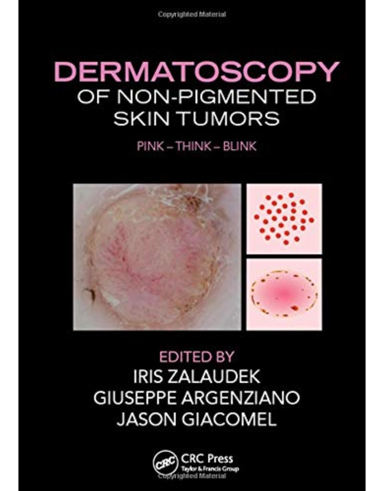
Dermatoscopy of Non-Pigmented Skin Tumors
Livrare gratis la comenzi peste 500 RON. Pentru celelalte comenzi livrarea este 20 RON.
Disponibilitate: La comanda in aproximativ 4 saptamani
Editura: CRC Press
Limba: Engleza
Nr. pagini: 158
Coperta: Hardcover
Dimensiuni: 184 x 261 x 15 mm
An aparitie: 28 Aug 2015
Although many skin lesions are pigmented, Dermatoscopy of Non-pigmented Skin Tumors: Pink - Think - Blink addresses non-pigmented lesions, which may be more difficult to diagnose. It discusses dermatoscopy not only as a reliable tool for diagnosis, but also for the monitoring of treatment outcomes following topical therapy.
The clinical diagnosis of non-pigmented skin lesions is one of the most challenging in the daily routine. To arrive at a correct diagnosis or at least an adequate management plan the clinician needs to collect many pieces of information and put them together like a puzzle. Illustrated with nearly 200 color clinical and dermatoscopic photographs, this book is an invaluable guide for clinicians striving to solve the diagnostic puzzle and correctly identify non-pigmented lesions. "
Table of Contents:
SECTION I TECHNICAL ASPECTS
1 Physics of polarized and nonpolarized dermoscopy and digital photography
Key features
Key management
References
2 Instrument-dependent criteria
Key points
Key feature
Key management
References
3 Metaphoric and descriptive language in dermoscopy: Lessons from the cognitive sciences
Introduction: Metaphors in dermatology and dermoscopy
Descriptive terminology in dermoscopy
Schematic illustration
Visual metaphors
Synthesis: Seeking a clear, effective terminology in dermoscopy
Key features
Key management
References
4 How to perform dermoscopy of non-pigmented skin lesions
Key features
Key management
Flowchart for diagnostic procedure
Key feature
Key management
SECTION II DERMATOSCOPY OF NON-PIGMENTED LESIONS
5 How to assess a given non-pigmented lesion
Key features
Key management
References
6 Clinical assessment
Key features
Key management
7 Vascular morphologies
Key features
Key management
References
8 Vascular arrangements
Key features
Key management
Reference
9 Specific patterns
Key features
Key management
References
10 Dermatoscopic clues in non-pigmented lesions
Summary and key features
Introduction
Deciding whether a lesion is raised
White clues
White lines
Surface keratin, dermatoscopic white circles, and structureless areas in raised lesions
Other “non-vessel” clues to diagnosis
Vessel pattern analysis
Benign vascular patterns
Polymorphous vessels including a pattern of dots
Other vessel patterns
Flat lesions
Raised lesions
Combination of vessel clues with other non-vessel clues
References
11 The influence of tumor thickness on the vascular morphologies
Key features
Key management
References
SECTION III SPECIFIC DERMATOSCOPIC PATTERNS AND THEIR DIAGNOSTIC SIGNIFICANCE
12 Intradermal nevi (including Unna and Miescher types)
Key feature
Key management
Clinical characteristics
Dermoscopic features
Clinical management
References
13 Clark nevi in fair skin types
Key features
Key management
References
14 Spitz nevi
Key features
Key management
Introduction
Dermoscopy
References
15 Atypical Spitzoid neoplasms (atypical Spitz nevi, atypical Spitz tumors, Spitzoid melanoma): A clinicopathological update
Key features
Key management
References
16 Nevi in patients with Bap1 germ line mutation, red-hair polymorphism, and albinism
Key features
Key management
References
17 Amelanotic melanoma
Key features
Key management
Early amelanotic melanoma
Intermediate thickness amelanotic melanoma
Thick melanoma
References
18 Hypomelanotic melanoma
Key feature
Key management
References
19 Cutaneous melanoma metastases
Key features
Key management
References
20 Sebaceous hyperplasia
Short explanatory text
Key features
Key management
References
21 Seborrheic keratosis
Seborrheic keratosis
Summary
Clear cell acanthoma
Summary
Large cell acanthoma
References
22 Dermatofibromas
Key features
Key management
References
23 Angioma, pyogenic granuloma, angiokeratoma
Introduction
Cherry angioma
Pyogenic granuloma
Angiokeratoma
References
24 Benign adnexal lesions
Key features
Key management
Sebaceous lesions
Follicular lesions
Eccrine and apocrine lesions
References
25 Basal cell carcinoma
Key points
Introduction
Nodular basal cell carcinoma (nBCC)
Dermoscopic features
Superficial basal cell carcinoma (sBCC)
Dermoscopic features
Infiltrative basal cell carcinoma (iBCC)
Dermoscopic features
Fibroepithelial BCC
Dermoscopic features
Infundibulocystic BCC
Dermoscopic features
Basosquamous carcinoma
Dermoscopic features
Conclusion
References
26 Keratinocyte skin cancer
Introduction
Actinic keratosis
Bowen’s disease
Squamous cell carcinoma
References
27 Dermoscopy of cutaneous neuroendocrine (“Merkel cell”) carcinoma
Key features
Key management
Dermoscopy
Management
References
28 Malignant vascular, adnexal, and fibrous tissue tumors
Key feature
Key management
Malignant vascular tumors
Kaposi sarcoma (KS)
Angiosarcoma (AS)
Malignant tumors of the fibrous tissue
Dermatofibrosarcoma protuberans (DFSP)
Atypical fibroxanthoma (AFX) and malignant fibrous histiocytoma (MFH)
Malignant adnexal tumors
Sebaceous carcinoma
Trichilemmal carcinoma
Porocarcinoma
References
29 Clues for the differential diagnosis of inflammatory lesions from tumoral lesions
Key features
Discoid lupus erythematosus (DLE) versus actinic keratosis
Psoriasis versus superficial basal cell carcinoma (sBCC) and Bowen’s disease (BD)
Dermatitis versus mycosis fungoides (MF)
References
30 Dermoscopy for assessing surgical margins
Key feature
Key management
References
31 Dermoscopy in the treatment decision (surgical vs. topical)
Key features
Key management
References
32 Dermoscopy for treatment monitoring (recurrence vs. clearance)
Dermoscopic monitoring of medical treatments for nonmelanoma skin cancers
Key features
Key management
References
33 Diagnostic clues and management rules
Clues for diagnosing tumors showing dotted/coiled vessels
Clues for the management of nodular pink lesions
Clues for the differential diagnosis of flat non-pigmented tumors
Always obtain a histopathological diagnosis of a clinically and dermoscopically suspected diagnosis of pyogenic granuloma
Lesions with a polymorphous vascular pattern should always be considered suspicious and submitted for histopathological examination
If pigment is present, look at it first before assessing the vascular pattern
Clues to recognize melanoma mimicking nodular basal cell carcinoma
34 Confocal microscopy in the diagnosis and management of non-pigmented skin tumors (which, when, and when not)
Key features
Key management
Introduction
Best indications
Limitations
Conclusions
References
| An aparitie | 28 Aug 2015 |
| Autor | Iris Zalaudek, Giuseppe Argenziano, Jason Giacomel |
| Dimensiuni | 184 x 261 x 15 mm |
| Editura | CRC Press |
| Format | Hardcover |
| ISBN | 9781482237528 |
| Limba | Engleza |
| Nr pag | 158 |
-
-
-
-
 71200 lei 62000 lei
 -

-
-

-
-

Clientii ebookshop.ro nu au adaugat inca opinii pentru acest produs. Fii primul care adauga o parere, folosind formularul de mai jos.