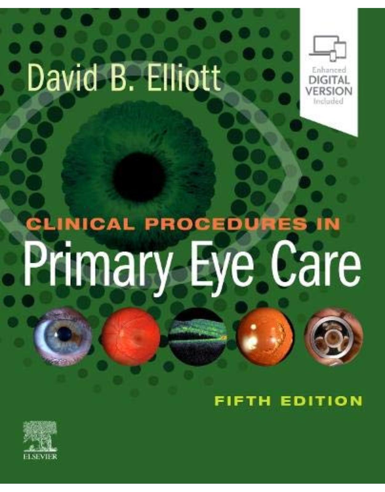
Clinical Procedures in Primary Eye Care
Livrare gratis la comenzi peste 500 RON. Pentru celelalte comenzi livrarea este 20 RON.
Disponibilitate: La comanda in aproximativ 4-6 saptamani
Editura: Elsevier
Limba: Engleza
Nr. pagini: 336
Coperta: Paperback
Dimensiuni: 19.69 x 1.27 x 24.13 cm
An aparitie: 1 May 2020
Description:
Well organized and easy to read, Clinical Procedures in Primary Eye Care, 5th Edition, takes an accessible, step-by-step approach to describing the commonly used primary care procedures that facilitate accurate diagnosis and effective patient management. This practical, clinically-focused text offers succinct descriptions of today's most frequently encountered optometric techniques supported by research-based evidence. You’ll find essential instructions for mastering the procedures you need to know, including recent technical advances in the field.
Discusses technical advances that are dramatically altering optometry: Optical Coherence Tomography (OCT) and ultra-wide field imaging (optomaps).
Presents outstanding new digital images of OCT cases and optomaps for a wide variety of conditions of the central and peripheral retina.
Focuses on evidence-based optometry; all procedures include a section that reviews when and how the procedure should be measured and uses clinical wisdom in addition to research-based evidence.
Presents new digital images of normal variations of the eye – crucial visual support for understanding what is normal and what is disease.
Helps you clearly visualize procedures and eye disorders through full-color photographs, diagrams, and video clips.
Provides fully revised print on dry eye assessment based on the latest international Dry Eye Workshop (DEWS) guidelines.
Features coverage of changes in the eye due to high myopia, and expounds the need for myopia control techniques.
Offers extensive material online to enhance learning: video clips, interactive testing sections, additional photographs, and more.
Table of Contents:
1. Evidence-based eye examinations
1.1 Several tests are widely used because of tradition and not evidence
1.2 Tips on reviewing the evidence base for clinical tests
1.3 “Screen everybody, don’t miss disease” vs. “what about false positives?”
1.4 Should I perform a database, system, and/or problem-oriented eye examination?
1.5 The evidence to support routine dilated fundus examinations is limited
1.6 Lessons to learn from the Honey Rose case
References
2. Communication skills
2.1 Turning anxious patients into satisfied ones
2.2 The importance of recording
2.3 Case history-taking
2.4 The 5 Rs: Providing diagnoses and management plan information
2.5 Giving bad news
2.6 What should a good referral letter include?
References
3. Assessment of visual function
3.1 Distance visual acuity
3.2 Reading (or near) acuity
3.3 Contrast sensitivity
3.4 Congenital colour vision deficiency
3.5 Acquired colour vision deficiency
3.6 Central visual field (24°) testing
3.7 Central visual field (10°), including Amsler
3.8 Peripheral visual field (30° to 60°)
3.9 Visual field assessment for drivers
3.10 Simple visual field assessments
3.11 Stereopsis
References
4. Refraction and prescribing
4.1 Focimetry (vertometry or lensometry)
4.2 Interpupillary distance
4.3 Phoropters are standard, but when are trial frames better?
4.4 Objective refraction
4.5 Monocular subjective refraction
4.6 Best vision sphere (maximum plus to maximum visual acuity)
4.7 Best vision sphere (plus/minus technique)
4.8 Duochrome (or bichromatic) test
4.9 Assessment of astigmatism
4.10 Binocular balancing of accommodation
4.11 Binocular subjective refraction
4.12 Cycloplegic refraction
4.13 The reading addition
4.14 Myopia control
4.15 Guidelines for prescribing glasses
References
5. Contact lens assessment
5.1 Contact lens fitting
5.2 Pre-fitting case history
5.3 Determining contact lens diameter
5.4 Corneal topography
5.5 Assessment of ocular physiology
5.6 Soft contact lens fitting
5.7 Selecting a soft trial lens
5.8 Assessment of the soft lens fit
5.9 Toric soft lens fitting
5.10 Presbyopic soft lens fitting
5.11 Fitting corneal RGP contact lenses
5.12 Scleral RGP lenses
5.13 Management of myopia progression
5.14 Patient instruction for contact lens care
5.15 Contact lens aftercare
References
6. Assessment of binocular vision and accommodation
6.1 Adaptations when testing younger patients
6.2 The cover test: Detection and assessment of strabismus and heterophoria
6.3 Other tests for the detection and measurement of strabismus
6.4 Motility test: Detection and assessment of incomitant strabismus
6.5 Convergence assessment: Near point of convergence
6.6 Suppression testing: Worth 4-dot and mallett test
6.7 Stereopsis
6.8 Accommodation assessment
6.9 Further assessment of binocular vision and accommodation (‘Functional tests’)
6.10 Further assessment of binocular status in strabismus
6.11 Further assessment of heterophoria
6.12 Fusional reserves
6.13 Fixation disparity
6.14 The accommodative convergence to accommodation relationship
6.15 Further assessment of the relationship between convergence and accommodation
6.16 Further evaluation of incomitant strabismus
6.17 Assessment of eye movements
6.18 Making a final diagnosis and management plan
References
7. Ocular health assessment
7.1 Examination of the anterior segment and ocular adnexa
7.2 Tear film and ocular surface assessment
7.3 Assessment of the lacrimal drainage system
7.4 Anterior chamber angle depth estimation
7.5 Examination of the anterior chamber angle by gonioscopy
7.6 Pachymetry
7.7 Tonometry
7.8 Instillation of diagnostic drugs
7.9 Pupil light reflexes
7.10 Fundus examination, particularly the posterior pole
7.11 Optical coherence tomography
7.12 Fundus examination of the peripheral retina
References
8. Variations in appearance of the normal eye
8.1 Anterior eye variations
8.2 Anterior eye changes in older patients
8.3 Lens and vitreous variations
8.4 Lens and vitreous changes in older patients
8.5 Optic nerve head variations
8.6 Fundus variations
8.7 Fundus changes in older patients
8.8 Peripheral fundus variations
8.9 Peripheral fundus changes in older patients
8.10 Myopic eyes
References
9. Physical examination procedures
9.1 Blood pressure measurement
9.2 Carotid artery assessment
9.3 Lymphadenopathy in the head–neck region
References
Subject index
| An aparitie | 1 May 2020 |
| Autor | David B. Elliott PhD MCOptom FAAO |
| Dimensiuni | 19.69 x 1.27 x 24.13 cm |
| Editura | Elsevier |
| Format | Paperback |
| ISBN | 9780702077890 |
| Limba | Engleza |
| Nr pag | 336 |



Clientii ebookshop.ro nu au adaugat inca opinii pentru acest produs. Fii primul care adauga o parere, folosind formularul de mai jos.