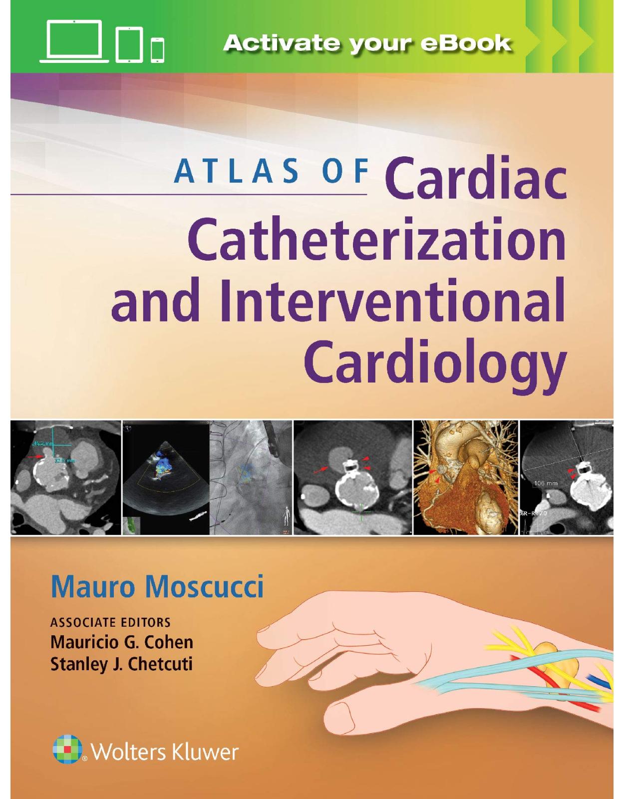
Atlas of Cardiac Catheterization and Interventional Cardiology: Practical Images for Diagnosis and Ablation
Livrare gratis la comenzi peste 500 RON. Pentru celelalte comenzi livrarea este 20 RON.
Disponibilitate: La comanda in aproximativ 4-6 saptamani
Editura: LWW
Limba: Engleza
Nr. pagini: 672
Coperta: Hardcover
Dimensiuni: 21.59 x 2.79 x 28.19 cm
An aparitie: 1 Dec. 2018
Description:
Comprehensive, current, and lavishly illustrated, Atlas of Interventional Cardiology thoroughly covers all of today’s cardiac catheterization and coronary interventional procedures, including catheterization and stenting techniques, angiography, atherectomy, thrombectomy, and more. Full-color illustrations and procedural videos guide you step by step through every interventional procedure you’re likely to perform.
Table of Contents:
1. Chapter 1 Integrated Imaging Modalities in the Cardiac Catheterization Laboratory
2. INTRODUCTION
3. CONCLUSION
4. Chapter 2 Complications of Percutaneous Coronary Intervention
5. CORONARY ARTERY DISSECTION
6. AORTIC AND GREAT VESSELS DISSECTION
7. CORONARY PERFORATION
8. AIR EMBOLISM
9. ARTERIAL ACCESS VASCULAR COMPLICATIONS
10. Thrombotic Complications
11. Atheroembolism
12. Hemorrhagic Complications
13. Chapter 3 Percutaneous Vascular Access: Transfemoral, Transseptal, Apical, and Transcaval Approach
14. INTRODUCTION
15. FEMORAL ACCESS
16. LARGE BORE ACCESS AND PRECLOSURE
17. ACCESS TO THE LEFT-SIDED HEART
18. TRANSAPICAL ACCESS
19. TRANSCAVAL ACCESS
20. Chapter 4 Radial Artery Approach
21. INTRODUCTION
22. CATHETERIZATION LABORATORY SETUP, PATIENT EVALUATION, AND PREPARATION
23. ARTERIAL ACCESS
24. RIGHT-SIDED HEART CATHETERIZATION
25. Postprocedure
26. VASCULAR ANATOMY
27. TRA INTERVENTIONS
28. Complications and Management
29. Chapter 5 Cutdown Approach: Femoral, Axillary, Direct Aortic, and Transapical
30. INTRODUCTION
31. FEMORAL ARTERY ACCESS (VIDEO 5.1)
32. AXILLARY ARTERY ACCESS (VIDEO 5.2)
33. DIRECT AORTIC ACCESS VIA UPPER MINISTERNOTOMY (VIDEO 5.3)
34. TRANSAPICAL LEFT VENTRICULAR ACCESS (VIDEO 5.4)
35. Chapter 6 Catheterization in Childhood and Adult Congenital Heart Disease
36. RIGHT VENTRICULAR OUTFLOW TRACT AND PULMONARY VALVE INTERVENTIONS
37. Hemodynamic Assessment
38. Indications and Risks of Intervention1,2a,b
39. Balloon Pulmonary Valvuloplasty
40. Right Ventricle to Pulmonary Artery Conduit Interventions
41. Transcatheter Pulmonary Valve Interventions
42. Subvalvar and Supravalvar Pulmonary Stenosis
43. Peripheral Pulmonary Stenosis Interventions
44. INTERVENTIONS FOR PULMONARY VEIN STENOSIS
45. Hemodynamic Assessment
46. Indications for Intervention
47. Key Aspects of Intervention
48. Short- and Long-Term Results
49. SHUNT CREATION
50. Balloon Atrial Septostomy
51. STENTING OF A DUCTUS ARTERIOSUS
52. Background
53. Indications1
54. Risks
55. Procedure
56. Short- and Long-Term Results
57. CLOSURE OF INTERATRIAL COMMUNICATIONS
58. Atrial Septal Defects, Patent Foramen Ovale Closure
59. Closure of Fontan Fenestrations or Baffle Leaks
60. CLOSURE OF VENTRICULAR SEPTAL DEFECTS
61. Background
62. Devices
63. Indications1
64. Risks
65. Procedure
66. PATENT DUCTUS ARTERIOSUS CLOSURE
67. Preprocedural Assessment
68. Indications and Risks of Intervention1,2a,b
69. Key Aspects of Intervention
70. Short- and Long-Term Results
71. CLOSURE OF EXTRACARDIAC SHUNTS
72. Assessment
73. Indications for Intervention1
74. Key Aspects of Intervention
75. Short- and Long-Term Results
76. AORTIC INTERVENTIONS
77. Coarctation of the Aorta
78. Midaortic Syndrome
79. Chapter 7 Pressure Measurements
80. INTRODUCTION
81. PRESSURE WAVEFORMS
82. Ventricular Pressure
83. Aortic Pressure
84. Atrial Pressure
85. Pulmonary Artery Wedge Pressure
86. PRESSURE TRACINGS IN VALVULAR AND NONVALVULAR HEART DISEASE
87. Chapter 8 Hemodynamics of Tamponade, Constrictive, and Restrictive Physiology
88. INTRODUCTION
89. TAMPONADE PHYSIOLOGY
90. CONSTRICTIVE PERICARDITIS
91. RESTRICTIVE CARDIOMYOPATHY
92. Chapter 9 Pitfalls in the Evaluation of Hemodynamic Data
93. INTRODUCTION
94. Chapter 10 Coronary Angiography and Cardiac Ventriculography
95. INTRODUCTION
96. CORONARY ANGIOGRAPHIC VIEWS
97. BYPASS ANGIOGRAPHY
98. CARDIAC VENTRICULOGRAPHY
99. Chapter 11 Coronary Anomalies
100. INTRODUCTION
101. ANOMALIES OF ORIGINATION AND COURSE
102. CORONARY FISTULAE
103. Chapter 12 Pulmonary Angiography
104. INTRODUCTION
105. ANATOMY OF THE PULMONARY ARTERY
106. TECHNICAL CONSIDERATIONS
107. Pulmonary Artery Catheterization
108. Injection Factors and Imaging Methods
109. Risks and Contraindications
110. INDICATIONS
111. Pulmonary Embolism
112. Chronic Pulmonary Embolism
113. Pulmonary Artery Aneurysms and Pseudoaneurysms
114. Pulmonary Artery Stenosis
115. Meandering Pulmonary Vein
116. Partial Anomalous Pulmonary Venous Connection
117. Retrieval of Pulmonary Artery Foreign Body
118. Chapter 13 Angiography of the Aorta and Peripheral Arteries
119. Introduction
120. ASCENDING AORTA
121. THORACIC AORTA
122. ABDOMINAL AORTA
123. DISTAL AORTA
124. LOWER EXTREMITY
125. Chapter 14 Myocardial and Coronary Blood Flow and Metabolism
126. BASIC PRINCIPLE OF FFR
127. Epicardial Coronary Artery Versus Microcirculation
128. Regulation of Coronary Blood Flow
129. Derivation and Experimental Basis for FFR
130. Core Concept of FFR
131. Coronary Pressure Remains Constant Throughout the Coronary Tree
132. Myocardial Perfusion Area Factored in by FFR
133. FFR as a Continuum of Risk
134. FFR Cutoff Point
135. FFR TECHNIQUE AND INDUCTION OF HYPEREMIA
136. Basic FFR Setup
137. Hyperemic Agents
138. Inducing Hyperemia With Adenosine
139. Limitations of IV Adenosine as Hyperemic Agent
140. FFR TECHNOLOGY
141. Piezoelectric Wires
142. Fiberoptic FFR Technology
143. Rapid Exchange Fiberoptic Microcatheter
144. FFR EVIDENCE
145. Intermediate Lesions Ideal for FFR Measurement
146. DEFER Trial
147. FAME Trial
148. FAME II Trial
149. R3F Trial
150. RIPCORD Study
151. ISIS Trial
152. HEMODYNAMIC ASSESSMENT OF SPECIFIC CORONARY LESIONS
153. Tandem Lesions or Serial Epicardial Lesions
154. Side Branch Evaluation Post-PCI
155. Collateral Circulation
156. Branch Vessel Disease
157. Proximal Versus Distal Coronary Stenoses
158. Multivessel CAD
159. Diffuse Atherosclerosis
160. Left Main Disease
161. Ostial Disease
162. PITFALLS OF FFR MEASUREMENTS
163. Chapter 15 Intravascular Imaging
164. Introduction
165. INTRAVASCULAR ULTRASOUND
166. Approaches to IVUS Imaging
167. Cross-sectional IVUS
168. Deviations
169. Image Orienation
170. Image Artifact
171. Cross-Sectional Ivus Measurements
172. Arterial Remodeling
173. Plaque
174. Coronary Artery Spasm
175. Perivascular Microvessels
176. Ivus Guidance For LMCA
177. OPTICAL COHERENCE TOMOGRAPHY
178. OCT Imaging Systems
179. Cross-sectional Format of OCT
180. Common OCT Image Artifacts (FIGURE 15.49)
181. OCT Assessment of Metal Stent Struts
182. Plaque Types by OCT (FIGURE 15.51)
183. Incomplete Stent Apposition (ISA)
184. Tissue Protrusion
185. Arterial Dissection
186. Thrombus and Ruptured Plaque
187. Cholesterol Crystals
188. Thin-cap Fibroatheroma (TCFA)
189. Stent Restenotic Tissue
190. Calcium Assessment
191. OCT Guidance for Severely Calcified Lesion
192. Microvessels
193. Peristrut Low-Intensity Area (PLIA)
194. Macrophage Infiltration
195. BRS on OCT
196. Spasm on OCT
197. Bifurcation Treatment and 3D OCT
198. SPECTROSCOPY
199. Basic Principles of Intravascular Spectroscopy (FIGURE 15.67)
200. Integrated Display and Quantification of Lipid Burden (FIGURE 15.68)
201. Histological Validation of Spectroscopy
202. Identification of Fibroatheroma with Calcification
203. Implication of NIRS-Detected Lipid-rich Plaque
204. Clinical Practice of Spectroscopy
205. EMERGING TECHNOLOGIES
206. Hybrid IVUS and OCT
207. High-definition (HD)-IVUS
208. Hybrid OCT Atherectomy
209. Co-registration of Angiography and Intravascular Imaging
210. Intracardiac Echocardiography (ICE)
211. ACKNOWLEDGMENTS
212. Chapter 16 Endomyocardial Biopsy
213. INTRODUCTION
214. Bioptomes
215. VASCULAR APPROACH
216. ECHOCARDIOGRAPHY-GUIDED BIOPSY
217. COMPLICATIONS
218. ENDOMYOCARDIAL BIOPSY IN THE EVALUATION OF TRANSPLANT REJECTION
219. ENDOMYOCARDIAL BIOPSY IN THE DIAGNOSIS AND MANAGEMENT OF CARDIOVASCULAR DISEASE
220. Chapter 17 Percutaneous Circulatory Support: Intra-Aortic Balloon Counterpulsation, Impella, Tandem Heart, and Extracorporeal Bypass
221. INTRODUCTION
222. LVAD AND AMCS
223. Interagency Registry for Mechanically Assisted Circulatory Support (INTERMACS)
224. AMCS Indications and Complications
225. THERAPEUTIC OBJECTIVES
226. Mechanical Circulatory Support Systems (MCS)
227. CF-AMCS
228. PV Loop
229. Clinical Scenarios
230. INTRA-AORTIC BALLOON PUMP (IABP) CONFIGURATION AND FUNCTION
231. BCIS-1 AND IABP-SHOCK TRIALS
232. TANDEMHEART (CARDIACASSIST, INC., PITTSBURG, PA)
233. Clinical Trials
234. IMPELLA AXIAL PUMP CATHETER
235. Safety and Efficacy
236. RIGHT ATRIAL-TO-FEMORAL ARTERY CENTRIFUGAL FLOW PUMPS
237. VA-ECMO
238. RIGHT VENTRICULAR FAILURE (RVF)
239. RVF After LVAD Implantation
240. RVF After Left Ventricular Support
241. Pulmonary Artery Pulsatility Index (PAPi) prior and immediately after LVAD
242. Biventricular Failure Support Options
243. Chapter 18 Percutaneous Transluminal Coronary Angioplasty
244. INTRODUCTION
245. GUIDE CATHETERS
246. GUIDE WIRES
247. PTCA BALLOONS
248. NEW TECHNOLOGY
249. ROLE OF PTCA IN THE MANAGEMENT OF CORONARY ARTERY DISEASE
250. Chapter 19 Atherectomy, Thrombectomy, and Distal Protection Devices
251. ATHERECTOMY, THROMBECTOMY, AND DISTAL PROTECTION DEVICES
252. Atherectomy
253. Thrombectomy
254. Embolic Protection
255. Chapter 20 Coronary Stenting
256. INTRODUCTION
257. LIMITATIONS OF PERCUTANEOUS TRANSLUMINAL CORONARY ANGIOPLASTY (PTCA)
258. DEFINITIONS OF RESTENOSIS
259. BALLOON ANGIOPLASTY AND STENTING
260. Chapter 21 Percutaneous Interventions for Valvular Heart Disease
261. Transcatheter Aortic Valve Interventions
262. TRANSCATHETER AORTIC VALVE REPLACEMENT: EDWARDS SAPIEN VALVE
263. Edwards Sapien Valve and Commander Delivery System (Figure 21.1)
264. Preprocedural Planning Using MDCT (Figure 21.2)
265. Hemodynamic Tracings
266. Assessment of Transcatheter Heart Valves (THV)
267. TRANSCATHETER AORTIC VALVE REPLACEMENT: MEDTRONIC COREVALVE EVOLUT
268. Measurements and Assessments
269. Hemodynamic Tracings
270. TRANSCATHETER VALVE-IN-VALVE INTERVENTIONS
271. AORTIC BIOPROSTHETIC VALVE-IN-VALVE: EDWARDS SAPIEN VALVE
272. AORTIC AND MITRAL VALVE-IN-VALVE: EDWARDS SAPIEN VALVE
273. TRICUSPID BIOPROSTHETIC VALVE-IN-VALVE: EDWARDS SAPIEN VALVE
274. Echocardiography
275. AORTIC BIOPROSTHETIC VALVE-IN-VALVE: MEDTRONIC COREVALVE EVOLUT
276. TRANSCATHETER AORTIC VALVE REPLACEMENT FOR SEVERE AORTIC INSUFFICIENCY AFTER LEFT VENTRICULAR ASSIST DEVICE PLACEMENT: MEDTRONIC COREVALVE EVOLUT
277. PERCUTANEOUS MITRAL BALLOON VALVULOPLASTY FOR MITRAL STENOSIS
278. PERCUTANEOUS TREATMENT FOR MITRAL REGURGITATION: THE MITRAL CLIP
279. TEE
280. Fluoroscopic Views
281. Chapter 22 Interventions for Adult Structural Heart Disease
282. PERCUTANEOUS REPAIR OF PARAVALVULAR LEAKS
283. PERCUTANEOUS CLOSURE OF VENTRICULAR SEPTAL DEFECTS
284. PERCUTANEOUS CLOSURE OF PATENT FORAMEN OVALE
285. PERCUTANEOUS CLOSURE OF PATENT DUCTUS ARTERIOSUS
286. PERCUTANEOUS PULMONARY VEIN STENOSIS INTERVENTION
287. TRANSCATHETER LEFT ATRIAL APPENDAGE OCCLUSION: WATCHMAN DEVICE
288. ENDOVASCULAR TREATMENT OF SAPHENOUS VEIN GRAFTS ANEURYSMS
289. Chapter 23 Peripheral Interventions
290. INTRODUCTION
291. ARTERIAL INTERVENTIONS
292. Carotid Arteries
293. Subclavian Arteries
294. Renal Arteries
295. Mesenteric Arteries
296. NORMAL LOWER EXTREMITY ARTERIAL ANATOMY (FIGURE 23.23)
297. Aortoiliac Occlusive Disease
298. Superficial Femoral and Popliteal Arteries
299. Infrapopliteal Arteries
300. VENOUS INTERVENTIONS
301. Inferior Vena Cava Filter
302. Chronic Inferior Vena Cava Occlusion
303. Acute and Subacute Inferior Vena Cava Occlusion
304. Chapter 24 Thoracic Aortic Endovascular Repair
305. INTRODUCTION
306. POSSIBLE CONFLICT OF INTEREST
307. ACKNOWLEDGMENTS
308. Chapter 25 Percutaneous Epicardial Techniques
309. ANTERIOR AND INFERIOR APPROACH
310. FLUOROSCOPIC NAVIGATION OF THE EPICARDIAL SPACE
311. Index
| An aparitie | 1 Dec. 2018 |
| Autor | Mauro Moscucci , Mauricio G Cohen MD FACC FSCAI, Stanley J Chetcuti MD |
| Dimensiuni | 21.59 x 2.79 x 28.19 cm |
| Editura | LWW |
| Format | Hardcover |
| ISBN | 9781451195163 |
| Limba | Engleza |
| Nr pag | 672 |




Clientii ebookshop.ro nu au adaugat inca opinii pentru acest produs. Fii primul care adauga o parere, folosind formularul de mai jos.