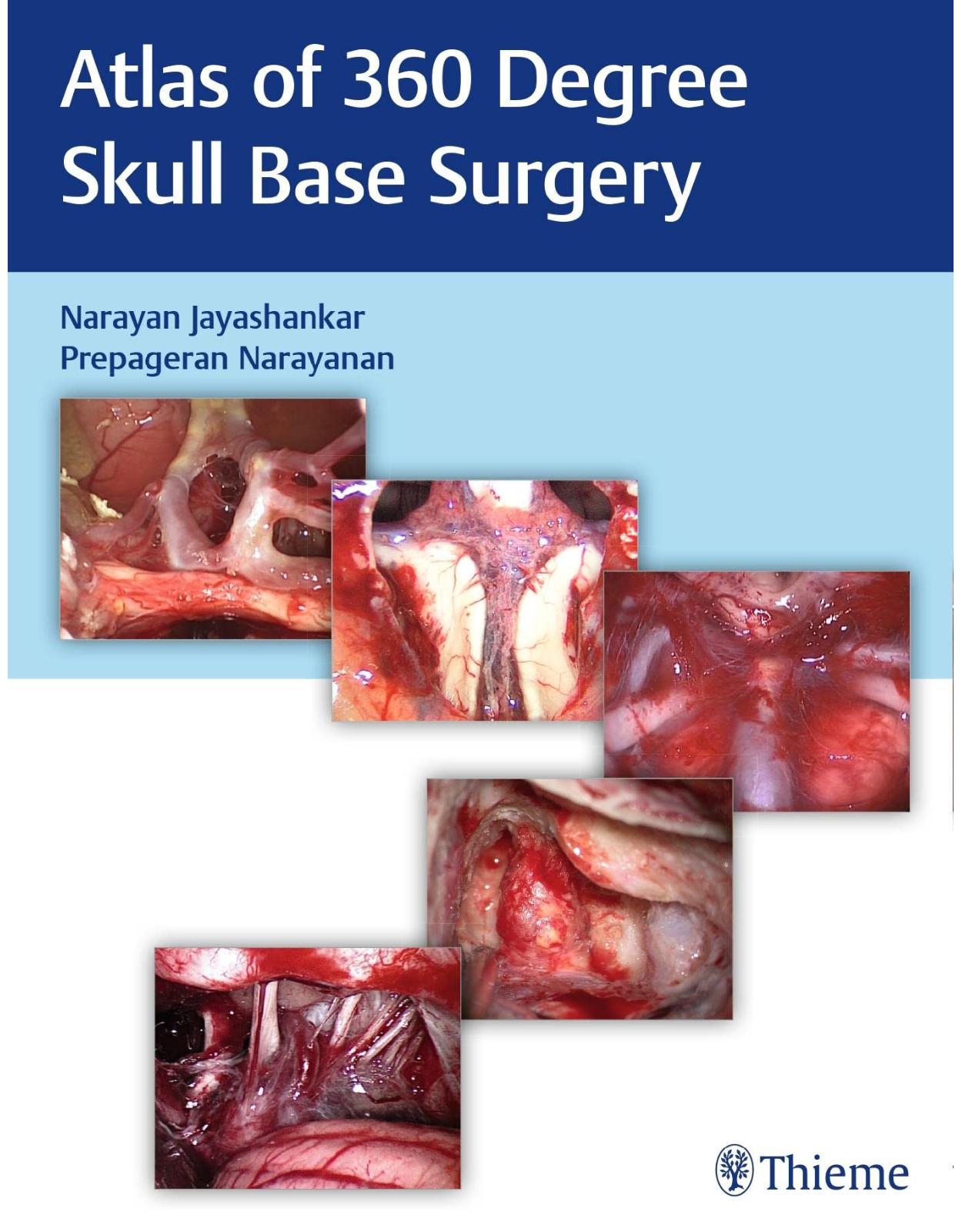
Atlas of 360 Degree Skull Base Surgery
Livrare gratis la comenzi peste 500 RON. Pentru celelalte comenzi livrarea este 20 RON.
Disponibilitate: La comanda in aproximativ 4-6 saptamani
Editura: Thieme
Limba: Engleza
Nr. pagini: 1064
Coperta: Hardcover
Dimensiuni: 22.6 x 4.5 x 28.7 cm
An aparitie: 31 Dec. 2021
Skull base surgery has evolved over the last decade into a symbiotic combination of cutting-edge technology and surgical skills. From the ORL perspective, skull base is usually performed by three separate teams, namely, rhinologist who performs the endoscopic transnasal skull base surgery, neurotologist who performs posterolateral skull base, and head and neck surgeon who performs the open skull base approaches. These traditional division, although still very much applicable in most countries, is now being challenged by some group of surgeons who perform the entire skull base surgery.
The editors of this surgical atlas belong to this group who performs the entire range of skull base as the surgical skills and instruments are interchangeable. Current fellowship includes exposure to the entire range of skull base, thus pioneering a younger generation of skull base surgeons who inherit the same versatility and technical capability of performing the entire range of skull base surgery. Many colleagues around the globe practice a single corridor of approaches. However, we believe in practicing 360-degree skull base, thus the need for this unique atlas that encompasses the entire skull base.
Key Features:
- Step-by-step description of procedures with detailed photographs
- More than 2,000 high-definition photographs to enhance the understanding of the text
- Detailed description of tips, tricks, and pitfalls to be avoided is covered in the atlas.
- Complications and their management, reconstruction of skull base defects, and rehabilitation chapters
- Comprehensive overview of pituitary tumors, craniopharyngioma, meningioma, craniovertebral junction pathologies, and chordoma
Table of Contents:
Cover
Title Page
Dedication
Contents
Foreword
Preface
About the Authors
Contributors
Abbreviations
Section I: Pituitary Gland Tumors
1. Pathologies of the Sellar Region
2. Endoscopic Transnasal Approach for Pituitary Tumors
3. Endoscopic Endonasal Surgeries for Pituitary Macroadenoma
4. Endoscopic Endonasal Revision Surgery for Pituitary Adenoma
5. Endoscopic Endonasal Surgery for Functioning Pituitary Adenoma
6. Endoscopic Endonasal Surgery for Cystic Lesions of the Sella
Section II: Supra-sellar Region
7. Craniopharyngioma
8. Endoscopic Endonasal Transsphenoidal Transplanum Transtuberculum Approach
9. Endoscopic Endonasal Surgeries for Craniopharyngioma
10. Microsurgical Approach for Suprasellar Lesions
11. Sellar, Parasellar, and Suprasellar Meningioma
12. Tailored Endonasal Endoscopic Approaches for Meningioma Involving the Planum Sphenoidale and Tuberculum Sellae
13. Pterional Craniotomy and Resection of Tuberculum Sellae Meningioma
Section III: Transcribriform Approach
14. Endoscopic Resection of Sinonasal Tumors with Endonasal Craniectomy
Section IV: Clival Region
15. Skull Base Chordoma
16. Transclival Approach to the Skull Base and Posterior Fossa
17. Endoscopic Endonasal Surgeries for Clival Chordomas
Section V: Craniocervical Junction
18. Pathologies of the Craniocervical Junction
19. Endonasal Endoscopic Surgeries of the Craniocervical Junction
Section VI: Orbital Region
20. Approach to Orbital Lesions: Intraconal and Extraconal
21. Endonasal Endoscopic Surgeries of the Orbit
Section VII: Orbital Apex and Optic Nerve
22. Pathologies of the Orbital Apex and Optic Nerve
23. Endonasal Endoscopic Surgeries of the Orbit Apex and Optic Nerve
Section VIII: Infratemporal Fossa and Parapharyngeal Space
24. Endoscopic Transpterygoid Approaches to the Coronal Plane
25. Endonasal Endoscopic Surgeries of Lesions Occupying the Infratemporal Fossa and the Parapharyngeal Space
26. Management of Juvenile Nasopharyngeal Angiofibroma
Section IX: Petrous Region
27. Pathologies of the Petrous Apex
28. Endonasal Endoscopic Surgeries for Petrous Apex Lesions
29. Endoscopic Petroclival Approach
Section X: Meckel’s Cave
30. Meckel’s Cave Lesions
31. Endonasal Endoscopic Surgeries at Meckel’s Cave
32. Trigeminal Schwannomas
Section XI: Cerebrospinal Fluid Rhinorrhea
33. Endoscopic Management of Cerebrospinal Fluid Rhinorrhea
34. Endonasal Endoscopic Surgeries for Cerebrospinal Fluid Leakage
35. Open Surgical Management of Encephaloceles
Section XII: Reconstruction in Endoscopic Skull Base
36. Principles and Techniques of Reconstruction in Skull Base Defects
Section XIII: Cavernous Sinus
37. Transcavernous and Anterior Transpetrous Approaches
38. Cavernous Sinus Surgery
Section XIV: Sinonasal Tumors
39. Sinonasal Tumors
40. Endonasal Endoscopic Management of Sinonasal Tumors
Section XV: Skullbase Fibro-osseous Pathologies
41. Skull Base Fibro-osseous Pathologies
42. Endoscopic Management of Skull Base Fibro-osseous Pathologies
43. Combined Open and Endonasal Endoscopic Management of Skullbase Fibro-osseous Pathologies
Section XVI: Complications
44. Complications in Endoscopic Skull Base Surgery
Section XVII: Basics in Otology
45. Cortical Mastoidectomy
46. Posterior Tympanotomy and Extended Posterior Tympanotomy
47. Facial Nerve Anatomy and Relationships
Section XVIII: Translabyrinthine Approach
48. The Enlarged Translabyrinthine Approach
49. Translabyrinthine Approach for the Resection of a Large Vestibular Schwannoma
Section XIX: Approaches Through the Otic Capsule
50. Modified Transcochlear Approach Type A
51. Transotic Approach
52. Modified Transcochlear Approach Type B
53. Modified Transcochlear Approach Type C
Section XX: Retrolabyrinthine Approach
54. Petrous Apex Cholesterol Granuloma Excision—Transmastoid Retrolabyrinthine Approach
Section XXI: Presigmoid Approach
55. Presigmoid Retrolabyrinthine Approach
Section XXII: Management of Glomus Tumors/Approaches to Jugular Foramen
56. Glomus Tympanicum Tumors
57. Management of Class B1 and B2 Temporal Bone Paragangliomas (Glomus Tumors)
58. Management of Class B3 Temporal Bone Paragangliomas (Glomus Tumors)
59. Management of Type C Glomus Tumors: Infratemporal Fossa Type A Approach for Glomus Jugulare Tumors
60. Petro-Occipital Transsigmoid Approach
Section XXIII: Approach to Foramen Magnum Tumors
61. Far and Extreme Lateral Transcondylar Approach to the Foramen Magnum
Section XXIV: Surgeries for Vertigo
62. Endolymphatic Sac Decompression
63. Transmastoid Labyrinthectomy
64. Vestibular Neurotomy by Retrosigmoid Approach
Section XXV: Microvascular Decompression
65. Endoscope-Assisted Retrosigmoid Approach in Neurovascular Decompression
Section XXVI: Retrosigmoid Approach
66. Retrosigmoid Approach
67. Retrosigmoid Craniotomy with Stereotactic Supratentorial Cerebrospinal Fluid Diversion
Section XXVII: Middle Cranial Fossa
68. Middle Cranial Fossa Approach
69. Extended Middle Cranial Fossa Approach
Section XXVIII: Otogenic Infections and Tumors
70. Single-Stage Transmastoid Drainage of Otogenic Intracranial Abscess
71. Subtotal Petrosectomy for Cochlear Implantation
72. Lateral Temporal Bone Resection
73. Facial Neuroma
Section XXIX: Rehabilitative Surgery
74. Hearing Restoration in Skull Base Surgery
75. Reanimation of the Paralyzed Face
76. Rehabilitation of Lower Cranial Palsies after Skull Base Surgeries
Index
| An aparitie | 31 Dec. 2021 |
| Autor | Narayan Jayashankar, Prepageran Narayanan |
| Dimensiuni | 22.6 x 4.5 x 28.7 cm |
| Editura | Thieme |
| Format | Hardcover |
| ISBN | 9789390553136 |
| Limba | Engleza |
| Nr pag | 1064 |


Clientii ebookshop.ro nu au adaugat inca opinii pentru acest produs. Fii primul care adauga o parere, folosind formularul de mai jos.