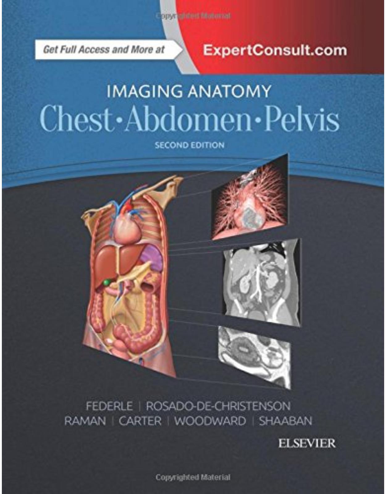
Imaging Anatomy: Chest, Abdomen, Pelvis, 2nd Edition
Produs indisponibil momentan. Pentru comenzi va rugam trimiteti mail la adresa depozit2@prior.ro sau contactati-ne la numarul de telefon 021 210 89 28 Vedeti mai jos alte produse similare disponibile.
Disponibilitate: Acest produs nu este momentan in stoc
Editura: Elsevier
Limba: Engleza
Nr. pagini: 1192
Coperta: Hardback
Dimensiuni: 222 X 281 mm
An aparitie: 2017
Description:
Designed to help you quickly learn or review normal anatomy and confirm variants, Imaging Anatomy: Chest, Abdomen, Pelvis provides detailed views of anatomic structures in successive imaging slices in each standard plane of imaging. Axial, coronal, sagittal, and 3D reconstructions accompany highly accurate and detailed medical drawings, assisting you in making an accurate diagnosis. Comprehensive coverage of the chest, abdomen, and pelvis, combined with an orderly, easy-to-follow structure, make this unique title unmatched in its field.
Includes all relevant imaging modalities, 3D reconstructions, and highly accurate and detailed medical drawings that illustrate the fine points of the imaging anatomy
Depicts common anatomic variants and covers common pathological processes as a part of its comprehensive coverage
Provides a detailed overview of airway and interstitial network anatomy—the basis for understanding and diagnosing interstitial lung disease
Features representative pathologic examples to highlight the effect of disease on human anatomy
Includes plain radiography, the latest generation of multi-planar advanced cross-sectional MR and CT, ultrasound for pelvis/renal/liver/gallbladder, barium for GI tract, and much more
Offers state of the art, detailed pelvic floor imaging and perianal/perirectal fistula imaging using high-resolution CT and MR, including 3T MR
Table of Contents
Copyright
Dedications
Additional Contributing Authors
Preface
Acknowledgments
Sections:
SECTION 1: CHEST
Chapter 1: Chest Overview
TERMINOLOGY
GENERAL ANATOMY AND FUNCTION
CHEST RADIOGRAPHY
COMPUTED TOMOGRAPHY
MAGNETIC RESONANCE IMAGING
ANGIOGRAPHY
OTHER CHEST IMAGING MODALITIES
Chest Overview
PA Chest Radiography
Lateral Chest Radiography
PA Chest Radiography, Positioning and Collimation
PA Chest Radiography
Left Lateral Chest Radiography, Positioning and Collimation
Left Lateral Chest Radiography
AP Chest Radiography, Positioning and Collimation
Portable AP Chest Radiography, Trauma and Intensive Care
PA and AP Chest Radiography
Inspiratory and Expiratory Chest Radiography
Lateral Decubitus Chest Radiography, Positioning and Collimation
Apical Lordotic Chest Radiography, Positioning
Radiographic Densities
Radiographic Densities, Medical Devices
Silhouette Sign
Anatomic Localization With Orthogonal Radiography
Decubitus Radiography for Evaluation of Complex Pleural Disease
Lordotic Chest Radiography for Evaluation of Apical Lesion
Silhouette Sign, Left Lower Lobe Airspace Disease
Silhouette Sign, Middle Lobe Airspace Disease
Cross-sectional Anatomy
Axial CT, Window and Level Settings
NECT and CECT
Section Thickness and HRCT
Supine and Prone HRCT
Inspiratory and Expiratory HRCT
CT Maximum- and Minimum-Intensity Projections
Coronal and Sagittal CT Angiography
CT Volume Rendering
Axial, Coronal, and Sagittal MR
CT, Acute Pulmonary Emboli
Angiography and Thrombolysis, Acute Pulmonary Emboli
Radiography and CT, Lung Cancer
CT and FDG PET/CT, Lung Cancer
Radiography and Ventilation-Perfusion Scintigraphy
CT and Ultrasonography, Empyema
Chapter 2: Lung Development
GENERAL CONCEPTS
EMBRYONIC STAGE (26 DAYS to 6 WEEKS)
PSEUDOGLANDULAR STAGE (6-16 WEEKS)
CANALICULAR STAGE (16-28 Weeks)
SACCULAR STAGE (28-36 WEEKS)
ALVEOLAR STAGE (36 WEEKS to 8 YEARS)
IMPORTANT FACTORS FOR NORMAL PRENATAL LUNG DEVELOPMENT
NEONATAL LUNG
DISORDERS OF LUNG IMMATURITY
DISORDERS RELATED TO TRACHEOBRONCHIAL MORPHOLOGY
DEVELOPMENTAL DISORDERS
Early Lung Development
Embryonic Stage
Embryonic Stage
Embryonic and Pseudoglandular Stages
Pseudoglandular Stage
Pseudoglandular and Canalicular Stages
Saccular and Alveolar Stages
Pseudoglandular, Canalicular, and Alveolar Stages
Neonatal Lung
Retained Fetal Lung Fluid Syndrome
Respiratory Distress Syndrome
Morphology and Orientation of Mainstem Bronchi
Embryonic Stage, Tracheal Atresia
Embryonic Stage, Tracheoesophageal Fistula and Esophageal Atresia
Embryonic Stage, Extralobar Sequestration
Embryonic Stage, Bronchogenic Cyst
Pseudoglandular Stage, Bronchial Atresia
Chapter 3: Airway Structure
GENERAL ANATOMY AND FUNCTION
AIRWAY STRUCTURE
FUNDAMENTAL UNITS OF LUNG STRUCTURE
COLLATERAL CHANNELS
IMAGING ANATOMIC CORRELATIONS
ANATOMY-RELATED IMAGING ABNORMALITIES
Overview of Airway Structure
Bronchi, Bronchioles, and Acini
Microscopic Structure of Trachea, Bronchi, and Bronchioles
CT Imaging anatomy of Large Airways
CT Imaging Anatomy of Large Airways
Inspiratory HRCT, Large Airway Morphology
Expiratory HRCT, Large Airway Morphology
Radiography, Large Airway Cartilage Calcification
CT, Large Airway Cartilage Calcification
Small Airway Structure and Secondary Pulmonary Lobule
Microscopic Structure of Small Airways
Graphic, Microscopic Structure of Alveoli
Microscopic Structure of Alveoli
Tree-in-Bud Opacities
Tree-in-Bud Opacities, Infectious Bronchiolitis
Centrilobular Emphysema and Bullous Disease
Paraseptal and Panlobular Emphysema
Lobular Low Attenuation, Constrictive Bronchiolitis
Radiography and CT, Acinar Nodules
Chapter 4: Vascular Structure
OVERVIEW OF PULMONARY VASCULAR STRUCTURE
GENERAL ANATOMY AND FUNCTION
PULMONARY ARTERIES
CAPILLARY NETWORK
PULMONARY VEINS
BRONCHIAL ARTERIES
BRONCHIAL VEINS
PULMONARY LYMPHATICS
VASCULAR STRUCTURE OF SECONDARY PULMONARY LOBULE
ANATOMY-RELATED IMAGING ABNORMALITIES
Arteries of Secondary Pulmonary Lobule
CT of Pulmonary Arteries
CT of Pulmonary Arteries
Microscopy of Pulmonary and Bronchial Arteries
CT of Pulmonary Arteries
Capillary-Alveolar Interface
Microscopic Features of Capillary-Alveolar Interface
CT Angiography of Pulmonary Veins
CT of Pulmonary Veins
Veins of Secondary Pulmonary Lobule
CT of Normal Secondary Pulmonary Lobule
Pulmonary Lymphatics
Lymphatics of Secondary Pulmonary Lobule
Random Nodules, Miliary Metastases, and Miliary Infection
Random Nodules, Miliary Infection
Perilymphatic Nodules, Sarcoidosis
Perilymphatic Nodules, Lymphangitic Carcinomatosis
Chapter 5: Interstitial Network
OVERVIEW OF PULMONARY INTERSTITIUM
INTERSTITIAL FIBER NETWORK
IMAGING NORMAL PULMONARY INTERSTITIUM
IMAGING OF INTERSTITIAL LUNG DISEASE
CHEST RADIOGRAPHY OF ABNORMAL PULMONARY INTERSTITIUM
HIGH-RESOLUTION CT OF ABNORMAL PULMONARY INTERSTITIUM
ANATOMY-RELATED IMAGING ABNORMALITIES
Axial and Peripheral Interstitium
Axial, Parenchymal, and Peripheral Interstitium
Microscopic Features of Normal Pulmonary Interstitium
Radiography of Normal Pulmonary Interstitium
Normal Pulmonary Interstitium on CT
Radiography, Interstitial Pulmonary Edema
CT, Interstitial Pulmonary Edema
Radiography and CT, Lymphangitic Carcinomatosis
Radiography and CT, Sarcoidosis
Radiography and CT, Idiopathic Pulmonary Fibrosis
Chapter 6: Lungs
TERMINOLOGY
ANATOMY
LOBES
SEGMENTS
ANATOMY-RELATED IMAGING ABNORMALITIES
Overview of Lungs
Surface Anatomy of Anterior and Posterior Lungs
Surface Anatomy of Lateral Lungs
Surface Anatomy of Medial Lungs
Surface Anatomy of Lungs on Radiography
Surface Anatomy of Lungs on CT
Lung Lobes
Lung Lobes
Radiography of Lung Lobes
Coronal and Sagittal CT of Lung Lobes
Axial CT of Lung Lobes
Axial CT of Lung Lobes
Segments of Right Lung
Segments of Right Lung
Segments of Left Lung
Segments of Left Lung
Axial CT of Lung Segments
Axial CT of Lung Segments
Perfusion Scintigraphy of Lungs
Scintigraphy, Lobar and Segmental Abnormalities
Accessory Azygos Fissure
Superior and Inferior Accessory Fissures
Radiography, Lobar Pneumonia
Radiography, Segmental Airspace Disease
Radiography, Consolidation in Right Upper Lobe
Radiography, Segmental Airspace Disease
Radiography, Lobar Atelectasis
Radiography, Lobar Atelectasis
Chapter 7: Hila
TERMINOLOGY
GENERAL ANATOMY AND FUNCTION
HILAR ANATOMY
RADIOGRAPHY OF HILA
CT OF HILA
HILAR LYMPH NODES
HILAR SIGNS
ANATOMY-RELATED IMAGING ABNORMALITIES
Overview of Hila
Right Hilum
Left Hilum
Radiography of Normal Hila
CT of Normal Hila
Radiography of Normal Hila
Graphic and Radiography of Normal Hila
Radiography of Normal Hila
CT of Normal Hila
Axial CT of Normal Hila
Axial CT of Normal Hila
Coronal CT of Normal Hila
Coronal CT of Normal Hila
Graphic, Sagittal CT, and Sagittal MR of Normal Right Hilum
Graphic, Sagittal CT, and Sagittal MR of Normal Left Hilum
Axial MR of Normal Hila
Coronal MR of Normal Hila
Hilum Overlay Sign
Hilum Convergence Sign
Differential Diagnosis of Hilar Enlargement
Differential Diagnosis of Hilar Enlargement
Abnormal Hilar Height
Hilar Calcification
Unilateral Hilar Enlargement, Pulmonic Stenosis
Unilateral Hilar Enlargement, Pulmonic Stenosis
Unilateral Hilar Enlargement and Thick Intermediate Stem Line, Lung Cancer
Thick Intermediate Stem Line, Pulmonary Edema
Chapter 8: Airways
TERMINOLOGY
NOMENCLATURE
OVERVIEW OF AIRWAY ANATOMY
TRACHEAL ANATOMY
RIGHT BRONCHIAL ANATOMY
LEFT BRONCHIAL ANATOMY
VARIANTS OF BRONCHIAL ANATOMY
IMAGING OF AIRWAYS
ANATOMY-RELATED IMAGING ABNORMALITIES
Overview of Airway Anatomy
Anterior View, Segmental Airways
Posterior View, Segmental Airways
Central Airways
Central Airways
Segmental Anatomy, Right Lung
CT, Right Lung Segmental Bronchi
CT, Right Lung Segmental Bronchi
CT, Right Lung Segmental Bronchi
Segmental Anatomy, Left Lung
CT, Left Lung Segmental Bronchi
CT, Left Lung Segmental Bronchi
CT, Left Lung Segmental Bronchi
Virtual Bronchoscopy
Bronchoarterial Ratio
Radiography, Bronchiectasis
CT, Bronchiectasis
CT, Tracheal Narrowing
CT, Airway Neoplasms
Chapter 9: Pulmonary Vessels
PULMONARY ARTERIES
PULMONARY VEINS
NOMENCLATURE OF PULMONARY ARTERIES AND VEINS
IMAGING OF PULMONARY ARTERIES
PULMONARY LYMPHATICS
ANATOMY-RELATED IMAGING ABNORMALITIES
Overview of Pulmonary Vessels
Radiography of Normal Pulmonary Vessels
Radiography of Pulmonary Trunk
Anatomy of Pulmonary Arteries and Veins
Angiography of Pulmonary Arteries
Axial CT of Pulmonary Arteries and Veins
Axial CT of Pulmonary Arteries and Veins
Angiography of Pulmonary Veins
Coronal CT of Pulmonary Vein Variants
Coronal CT of Pulmonary Arteries and Veins
Coronal CT of Pulmonary Arteries and Veins
Axial CT of Right Pulmonary Arteries
Axial CT of Right Pulmonary Arteries
Axial CT of Right Pulmonary Arteries
Axial CT of Left Pulmonary Arteries
Axial CT of Left Pulmonary Arteries
Axial CT of Left Pulmonary Arteries
Graphic and CT of Pulmonary Lymphatics and Intrapulmonary Lymph Nodes
CT of Pulmonary Lymph Nodes
Vascular Redistribution, Pulmonary Venous Hypertension
Pulmonary Artery Enlargement, Pulmonary Hypertension
Abnormal Vascular Connections, Arteriovenous Malformation
Abnormal Vascular Connections, Hepatopulmonary Syndrome
Abnormal Vascular Connections, Partial Anomalous Pulmonary Venous Return
Abnormal Vascular Connections, Scimitar Syndrome
Acute Pulmonary Thromboembolic Disease
Chronic Pulmonary Thromboembolic Disease
Pulmonary Embolism and Foreign Materials
Pulmonary Embolism and Foreign Materials
Chapter 10: Pleura
GENERAL ANATOMY AND FUNCTION
PARIETAL PLEURA
VISCERAL PLEURA
PLEURAL REFLECTIONS
INTERLOBAR FISSURES
ACCESSORY FISSURES
IMAGING
ANATOMY-RELATED IMAGING ABNORMALITIES
Overview of Pleural Anatomy
Coronal Section, Anatomy of Pleural Reflections
Sagittal Section, Anatomy of Pleural Reflections
Anatomy of Pleural Fissures
Radiography, Normal Pleura and Fissures
Radiography and CT, Normal Pleura and Fissures
CT, Standard Fissures
Radiography, Minor Fissure
CT, Minor Fissure
Coronal CT, Standard Fissures
Anatomy and CT, Incomplete Fissures
CT, Accessory Fissures
Anatomy and Radiography, Accessory Azygos Fissure
Radiography, Pneumothorax
Radiography and CT, Pleural Effusion
Radiography, Pseudotumor
CT, Loculated Pleural Effusion and Empyema
Radiography and CT, Pleural Plaques
Radiography and CT, Fibrothorax
Morphology and CT, Pleural Masses
Radiography and CT, Localized Fibrous Tumor
Radiography and CT, Solid Pleural Metastases
Radiography and CT, Malignant Pleural Mesothelioma
Chapter 11: Mediastinum
GENERAL ANATOMY
RADIOGRAPHIC ANATOMY
CT and MR Anatomy
MEDIASTINAL COMPARTMENTS
THYMUS
MEDIASTINAL LYMPH NODES
ANATOMY-RELATED IMAGING ABNORMALITIES
Overview of Mediastinum
Anatomy of Mediastinum and Paravertebral Regions
Anatomy of Mediastinal Nerves
Mediastinal Contours, Frontal Radiography and Coronal CT
Mediastinal Contours on Lateral Radiography and Sagittal CT
Aortopulmonary Window and Aortic-Pulmonary Stripe
Anterior Junction Line
Posterior Junction Line
Paratracheal Stripes
Azygoesophageal Recess
Pleuroesophageal Stripes
Preaortic Recess
Paravertebral Stripes
Retrosternal Stripe
Posterior Tracheal Stripe
Cross-Sectional Mediastinal Anatomy
Axial CT, Mediastinal Anatomy
Axial CT, Mediastinal Anatomy
Coronal MR, Mediastinal Anatomy
Mediastinal Compartments, Anatomic and Radiographic
CT Mediastinal Compartments, ITMIG
CT Mediastinal Compartments, ITMIG
Thymus
CT, Adult Thymus
Normal Pediatric Thymus, Radiography and CT
IASLC Lymph Node Map
IASLC Mediastinal Lymph Nodes
IASLC Mediastinal Lymph Nodes
IASLC Mediastinal Lymph Nodes
Approach to Mediastinal Abnormalities
Approach to Mediastinal Abnormalities
Approach to Mediastinal Abnormalities
Approach to Mediastinal Abnormalities
Chapter 12: Systemic Vessels
GENERAL ANATOMY AND FUNCTION
SYSTEMIC ARTERIES
SYSTEMIC VEINS
THORACIC LYMPHATICS
RADIOGRAPHY OF GREAT VESSELS
CT/MR OF GREAT VESSELS
ANATOMY-RELATED IMAGING VARIANTS AND ABNORMALITIES
Overview of Thoracic Systemic Vessels
Anatomy of Systemic Vessels
Anatomy of Aorta and Systemic Arteries
Anatomy of Systemic Veins
Anatomy of Systemic Veins
Radiography of Systemic Vessels
Radiography of Systemic Vessel Interfaces
Axial CT of Systemic Vessels
Axial CT of Systemic Vessels
Axial CT of Systemic Vessels
Coronal CT of Systemic Vessels
Coronal CT of Systemic Vessels
Coronal, Sagittal, and Volume-Rendered CT of Systemic Veins
Sagittal MR of Thoracic Aorta and Branches
Coronal and Sagittal MR of Azygos Vein
Radiography and CT of Left Superior Intercostal Vein
Aortography
Graphic and CT of Bronchial Arteries
Hypertrophied Bronchial Arteries
Anatomic Variants of Bronchial Arteries
Hypertrophied Bronchial Arteries
Anatomy of Thoracic Lymphatics
CT of Thoracic Duct
Aortic Enlargement, Aneurysm
Aortic Enlargement, Aneurysm
Aortography and CT of Bovine Aortic Arch
CT of Aberrant Left Vertebral Artery
Graphic and Radiography of Aberrant Right Subclavian Artery
CT of Aberrant Right Subclavian Artery
Right Aortic Arch, Non-Mirror Image Branching
Right Aortic Arch, Non-Mirror Image Branching
Double Aortic Arch
Persistent Left Superior Vena Cava
Persistent Left Superior Vena Cava
Persistent Left Superior Vena Cava
Persistent Left Superior Vena Cava, Right Superior Vena Cava
Azygos Continuation of Inferior Vena Cava
Persistent Left Superior Vena Cava and Hemiazygos Continuation of Inferior Vena Cava
Persistent Left Superior Vena Cava and Hemiazygos Continuation of Inferior Vena Cava
Chapter 13: Heart
GENERAL ANATOMY AND FUNCTION
CARDIAC SHAPE AND ORIENTATION
CARDIAC SURFACES
CARDIAC BORDERS (MARGINS)
CARDIAC SULCI OR GROOVES
CARDIAC CHAMBERS
CARDIAC SKELETON
CARDIAC VALVES
IMAGING
IDENTIFICATION OF MORPHOLOGIC RIGHT AND LEFT VENTRICLES
IDENTIFICATION OF MORPHOLOGIC RIGHT AND LEFT ATRIA
ANATOMY-BASED CARDIAC AND VALVULAR ABNORMALITIES
Overview of Heart
Anatomy of Heart Surfaces, Margins, and Sulci
Anatomy of Heart Surfaces, Margins and Sulci
Radiography of Heart
CT of Heart Surfaces and Borders
Anatomy of Anterior Cardiac Surface and Right Atrium
Anatomy of Right Ventricle
CT of Right Cardiac Chambers
CT of Right Cardiac Chambers
Anatomy of Posterior Cardiac Surface, Cardiac Base, and Left Atrium
Anatomy of Left Ventricle
CT of Left Cardiac Chambers
CT of Left Cardiac Chambers
Coronal CT of Heart
Coronal CT of Heart
Sagittal CT of Heart
Sagittal CT of Heart
CT of Right Atrium
CT of Left Atrium
CT 4-Chamber View
CT 2-Chamber View
CT Short-Axis View
MR Short-Axis View
Sagittal MR of Heart
Sagittal MR of Heart
Anatomy of Cardiac Skeleton and Heart Valves
Radiography of Prosthetic Aortic and Mitral Valves
Graphic and CT of Atrioventricular Valves
Anatomy, CT, and MR of Semilunar Valves
Anatomy of Tricuspid and Pulmonic Valves
CT of the Right Heart Valves
Anatomy of Left Heart Valves
Anatomy and CT of Left Heart Valves
CT and MR of Aortic Valve
CT of Mitral Valve
MR of Valve Function
MR of Valve Function
Radiography of Heart
Radiography and CT of Left Atrial Enlargement
Radiography and CT of Left Ventricular Aneurysm
Radiography and CT of Calcific Aortic Stenosis and Mitral Annulus Calcification
Chapter 14: Coronary Arteries and Cardiac Veins
TERMINOLOGY
ARTERIES
VEINS
ANATOMY IMAGING ISSUES
ANATOMIC VARIANTS AND CLINICAL IMPLICATIONS
Anterior View of Coronary Arteries
Posterior View of Coronary Arteries
Left Ventricular Segmentation
Coronary Artery Territories
Coronary Artery Territories
Catheter Coronary Angiography
Catheter Coronary Angiography
Coronary Arteries
Right Dominant Coronary Circulation
Left Dominant Coronary Circulation
Maximum-Intensity Projection of Coronary Arteries
Maximum-Intensity Projection of Coronary Arteries
Coronary Artery Anomalies
Single Coronary Artery From Right Coronary Sinus
Single Right Coronary Artery
Anomalous Course of Left Coronary Artery
Anterior View of Coronary Veins
Coronary Sinus
Double Superior Vena Cava
Biventricular Pacing
Chapter 15: Pericardium
ANATOMY OF PERICARDIUM
PERICARDIAL SPACE AND PERICARDIAL RECESSES
IMAGING OF NORMAL PERICARDIUM
ANATOMY-RELATED IMAGING ABNORMALITIES
Overview of Pericardium
Anatomy of Pericardium
Anatomy of Pericardium
Anatomy of Pericardial Recesses
Axial CT of Pericardium
Coronal and Sagittal CT of Pericardium
Axial and Coronal MR of Pericardium
Sagittal MR of Pericardium
CT of Pericardial Sinuses
CT of Pericardial Recesses
CT of Pericardial Recesses
CT of Pericardial Recesses
Congenital Absence of Pericardium
Pericardial Cyst
Pericardial Effusion
Pericardial Effusion
Pericardial Thickening and Calcification
Constrictive Pericarditis
Chapter 16: Chest Wall
GENERAL ANATOMY AND FUNCTION
SKELETAL STRUCTURES
MUSCLES
VESSELS
SOFT TISSUES
ANATOMIC REGIONS
IMAGING
ANATOMY-RELATED IMAGING ABNORMALITIES
Chest Wall Overview
Chest Wall Anatomy
Thoracic Inlet, Supraclavicular and Axillary Regions
Anatomy and MR, Intercostal Region
Axial CT, Normal Chest Wall
Axial CT, Normal Chest Wall
Axial MR, Normal Chest Wall
Axial MR, Normal Chest Wall
Coronal CT, Normal Chest Wall
Coronal CT, Normal Chest Wall
Coronal MR, Normal Chest Wall
Coronal MR, Normal Chest Wall
Radiography and CT, Sternum
Radiography and CT, Poland Syndrome
Radiography, Pectus Excavatum
CT, Pectus Excavatum
Radiography, Pectus Carinatum
Radiography, Kyphoscoliosis
CT, Chest Wall Infection
CT, Chondrosarcoma and Osteosarcoma
CT, Osseous Metastases
CT, Lipoma and Liposarcoma
SECTION 2: ABDOMEN
Chapter 17: Embryology of Abdomen
GROSS ANATOMY
18-Day Embryo (Lateral)
18-Day Embryo (Cross Section)
4-Week Embryo (Lateral)
4-Week Embryo (Cross Section)
Development of GI Tract (5 Weeks)
Development of GI Tract (6 Weeks)
Development of GI Tract (8 Weeks)
Development of Lesser Sac
Development of GI Tract (10-Week Fetus)
Development of GI Tract (16-Week Fetus)
Development of Hepatic Veins (4- to 6-Week Fetus)
Development of Hepatic Veins (8- to 12-Week Fetus)
Prenatal Abdominal Circulation
Postnatal Circulation
Peritoneal Reflections in Paracolic Gutters
Sagittal Section of Mesenteries and Peritoneal Cavity
Peritoneal Reflections and Spaces
Peritoneal Reflections and Spaces
Peritoneal Reflections and Spaces
Mesenteries and Peritoneal Spaces
Mesenteries and Peritoneal Spaces
Retroperitoneal Spaces and Planes
Retroperitoneal Spaces and Planes
Communication Among Extraperitoneal Spaces
Communication Among Extraperitoneal Spaces
Communication Among Extraperitoneal Spaces
Development of GU Tract
Development of GU Tract (5-Week Fetus)
Urinary Bladder Development
Development of GU Tract (20-Week Male Fetus)
Development of GU Tract (20-Week Female Fetus)
Kidney Ascent and Rotation (6 Weeks)
Kidney Ascent and Rotation (7 Weeks)
Kidney Ascent and Rotation (9 Weeks)
Chapter 18: Abdominal Wall
TERMINOLOGY
GROSS ANATOMY
CLINICAL IMPLICATIONS
Posterior Abdominal Wall Muscles
Anterior Wall Muscles
Anterior Abdominal Wall
Thoracolumbar Fascia
Axial CT, Abdominal Wall Muscles
Axial CT, Abdominal Wall Muscles
Axial CT, Abdominal Wall Muscles
Axial CT, Abdominal Wall Muscles
Axial MR, Abdominal Wall Muscles
Coronal CT, Abdominal Wall, Muscles & Vessels
Coronal CT, Abdominal Wall, Muscles & Vessels
Coronal CT, Abdominal Wall, Muscles & Vessels
Coronal CT, Abdominal Wall Muscles
Coronal CT, Abdominal Wall Muscles
Coronal CT, Abdominal Wall Muscles
Rectus Hemorrhage Into Perivesical Space
Pannus Simulating Ventral Hernia
Ventral Hernia, Incarcerated Causing Bowel Obstruction
Incisional Hernia Containing Colon
Inguinal Hernia Causing Small Bowel Obstruction
Spigelian Hernia Causing Colonic Obstruction
Spigelian Hernia Causing Colonic Obstruction
Lumbar Hernia
Chapter 19: Diaphragm
GROSS ANATOMY
ANATOMY IMAGING ISSUES
CLINICAL IMPLICATIONS
Abdominal Surface of Diaphragm
Normal Axial CT, Diaphragm and Crura
Normal Axial CT, Diaphragm and Crura
Normal Coronal CT, Diaphragm and Crura
Normal Sagittal CT, Diaphragm and Crura
Coronal and Axial CT, Diaphragm
Normal Diaphragm, Demonstration of Crura and GE Junction
Normal Diaphragm, Demonstration of Crura and GE Junction
Diaphragm and GE Junction in Multiple Planes
Median Arcuate Ligament Syndrome
Sagittal CT, Median Arcuate Ligament Syndrome
Variant, Diaphragm With Prominent Slips
Diaphragm Separating Pleural and Peritoneal Fluid Collections
Elevated Hemidiaphragm, Phrenic Nerve Injury
Coronal and Sagittal CT, Eventration of Diaphragm
Bochdalek Hernia
Diaphragm With Loose Crus and Hiatal Hernia
Large Hiatal Hernia
Traumatic Diaphragmatic Rupture
Chapter 20: Peritoneal Cavity
TERMINOLOGY
GROSS ANATOMY
ANATOMY IMAGING ISSUES
CLINICAL IMPLICATIONS
Lateral View of Mesenteries and Peritoneal Cavity
Lesser Sac and Peritoneal Recesses
Omentum and Peritoneal Reflections
Peritoneal Spaces and Reflections
Peritoneal Spaces and Reflections
Peritoneal Spaces and Mesenteries
Peritoneal Spaces and Mesenteries
Distended Peritoneal Cavity
Distended Peritoneal Cavity
Peritoneal Mesenteries
Peritoneal Recesses (Morison and Douglas)
Lesser Sac (Omental Bursa)
Umbilical Ligaments, Delineated by Ascites
Umbilical Folds (Ligaments)
Peritonitis With Loculated Ascites
Bacterial Peritonitis With Ascites and Thickened Peritoneum
Fibrosing Peritonitis Due to Peritoneal Dialysis
Peritoneal Carcinomatosis
Peritoneal Carcinomatosis
Peritoneal Carcinomatosis
Pseudomyxoma Peritonei
Chapter 21: Vessels, Lymphatic System and Nerves, Abdominal
TERMINOLOGY
GROSS ANATOMY
Coronal Abdominal Arteries
Internal Iliac Artery
CT Angiogram, Normal Aortic Branches
Normal Catheter Aortogram
Angiogram, Celiac Artery
Angiogram, Superior Mesenteric Artery
Angiogram, Inferior Mesenteric Artery
Angiogram, Hepatic Artery Variants
CT, Abdominal and Pelvic Arterial Vasculature
CT, Abdominal and Pelvic Arterial Vasculature
CT, Abdominal and Pelvic Arterial Vasculature
CT, Abdominal and Pelvic Arterial Vasculature
CT, Abdominal and Pelvic Arterial Vasculature
CT, Abdominal and Pelvic Arterial Vasculature
CT, Abdominal and Pelvic Arterial Vasculature
CT, Abdominal and Pelvic Arterial Vasculature
CT, Hepatic Artery Variants
CT, Hepatic Artery Variants
CT, Renal and Hepatic Artery Variants
CT, Celiac Artery Variants
Abdominal and Pelvic Arterial and Venous Vasculature
Abdominal and Pelvic Arterial and Venous Vasculature
Abdominal and Pelvic Arterial and Venous Vasculature
Abdominal and Pelvic Arterial and Venous Vasculature
Coronal Abdominal Veins
Inferior Vena Cava Duplication and Anomalies
Variation, Left-Sided Inferior Vena Cava (Transposition)
Variation, Left-Sided Inferior Vena Cava (Transposition)
Variation, Inferior Vena Cava Duplication
Variation, Inferior Vena Cava Duplication
Variation, Circumaortic Renal Vein
Variation, Azygous Continuation of Inferior Vena Cava and Polysplenia
Variation, Accessory Hepatic Vein
Variation, Interrupted Inferior Vena Cava
Coronal Lymph Nodes
Normal Lymphangiogram
Postlymphangiogram CT
Lymphadenopathy Due to Lymphoma
Autonomic Nerves and Ganglia of Abdomen
Neural Invasion by Pancreatic Cancer
Chapter 22: Esophagus
TERMINOLOGY
GROSS ANATOMY
IMAGING ANATOMY
ANATOMY IMAGING ISSUES
CLINICAL IMPLICATIONS
Esophagus From Pharynx to Stomach
Esophagus Sagittal Relations
Gastroesophageal Junction & Lymphatics System
Normal Impressions on Esophagus
Barium Esophagram, Esophageal A & B Rings
Normal Gastroesophageal Junction
Gastroesophageal Junction
Patulous Diaphragmatic Hiatus With Hernia
Esophageal B Ring, Hiatal Hernia
Stricture at B Ring (Schatzki Ring)
Cricopharyngeal Achalasia & Hiatal Hernia
“Feline” Esophagus
Paraesophageal Hernia
Paraesophageal Hernia
Esophageal Varices
Chapter 23: Gastroduodenal
GROSS ANATOMY
CLINICAL IMPLICATIONS
Frontal View of Stomach & Gastric Mucosa
Duodenum, Frontal & Internal Fold Pattern
Lymphatics of Stomach & Small Intestine
Arteries of Stomach and Duodenum
Portal Venous System
Normal Stomach
Upper GI, Normal Stomach & Duodenum
Axial CECT, Normal Gastroduodenal Anatomy
Axial CECT, Normal Gastroduodenal Anatomy
Normal Gastric & Duodenal Vasculature
Catheter & CT Angiography, Arterial Variations
Arterial Variations, Separate Origin of Left Gastric Artery
Brunner Gland Hyperplasia
Perforated Duodenal Ulcer
Perforated Duodenal Ulcer
Perforated Duodenal Ulcer, Tissue Planes
Perforated Gastric Ulcer
Gastric Adenocarcinoma
Gastric Adenocarcinoma
Gastric Carcinoma With Gastrocolic Ligament Tumor Extension
Other Gastric Masses
Duodenal Adenocarcinoma
Gastrointestinal Stromal Tumors of Stomach and Duodenum
Duodenal Compression, SMA Syndrome
Aortoenteric Fistula
Gastric Diverticulum
Large Duodenal Diverticulum
Chapter 24: Small Intestine
GROSS ANATOMY
ANATOMY IMAGING ISSUES
CLINICAL IMPLICATIONS
EMBRYOLOGY
Small Intestine Vessels
Jejunum
Ileum
Mesentery Outlined by Ascites
MR Enterography, Normal Jejunum & Ileum
Barium Study, Normal Small Intestine
Enteroclysis, Normal Small Intestine
Lymphoid Nodules
Catheter Angiogram, Superior Mesenteric Artery & Vein
CT Angiogram, Superior Mesenteric Artery & Vein
HIV/AIDS With Involvement of Mesentery & Small Bowel
Crohn Disease
Crohn Disease
Crohn Disease
Crohn Disease With Complex Fistulizing Disease
Small Bowel Ischemia, SMA occlusion
Small Bowel Ischemia, Venous Thrombosis
Small Bowel Ischemia & Infarction With Pneumatosis & Portal Venous Gas
Small Intestine Obstruction
Small Intestine Obstruction
Plain Radiographs, Malrotation
CT, Malrotation
Fluoroscopy, Malrotation
Midgut Volvulus
Congenital Duplication Cyst
Meckel Diverticulum
Small Bowel Tumors
Small Bowel Tumors
Small Bowel & Mesenteric Trauma
Chapter 25: Colon
GROSS ANATOMY
CLINICAL IMPLICATIONS
Colon & Its Mesentery
Arteries
Veins
Ileocecal Region
Lateral View, Rectosigmoid
Rectal Veins & Valves
Barium Enema, Normal Colon
Barium Enema, Normal Colon
Normal Ileocecal Valve
Normal Appendix
Superior Mesenteric Vessels
Inferior Mesenteric Vessels
Mesentery Outlined by Ascites
Mesentery Outlined by Ascites
Inferior Mesenteric Vessels, Ascites
Inferior Mesenteric Vessels, Ascites
Inferior Mesenteric Vessels, Ascites
MR, Rectum
Anal Sphincters
Anal Sphincters
Anal Sphincters
Anal Sphincters
Rectum and Anal Canal
Anal Sphincter Complex
Acute Appendicitis
Acute Appendicitis
Diverticulitis
Diverticulitis
Epiploic Appendagitis
Colon Polyps
Sigmoid Volvulus
Cecal Volvulus
Cecal Volvulus
Cecal Volvulus
Colon Cancer
Rectal Cancer With Metastases
Ischemic Colitis
Ischemic Colitis
Rectal Cancer
Rectal Cancer
Chapter 26: Spleen
GROSS ANATOMY
ANATOMY IMAGING ISSUES
CLINICAL IMPLICATIONS
EMBRYOLOGY
Spleen and Its Relations
Axial CT, Normal Spleen
Coronal CT, Normal Spleen
Normal Spleen
Normal Spleen
Heterogeneous Splenic Enhancement
Splenic Ligaments and Vessels
Splenic Ligaments and Vessels
Axial T2-Weighted MR, Normal Spleen
Axial T2-Weighted MR, Normal Spleen
Angiogram, Splenic Vessels
Splenic Trauma
Lymphoma
Lymphoma
Splenic Vein Occlusion, Cancer
Intrasplenic Pseudocyst
Acute Splenic Infarction
Splenic Infarction and Cyst
Chronic Splenic Infarction in Sickle Cell Disease
Enlarged Accessory Spleens
Accessory Spleen, Intrapancreatic
Wandering Spleen
Polysplenia Syndrome
Chapter 27: Liver
GROSS ANATOMY
CLINICAL IMPLICATIONS
Hepatic Visceral Surface
Hepatic Attachments and Relations
Hepatic Vessels and Bile Ducts
Axial CT, Normal Liver
Axial CT, Normal Liver
Axial CT, Normal Liver
Portal Venous System
Liver Segmental Anatomy
Liver Segmental Anatomy
Liver Segmental Anatomy
Liver Segmental Anatomy
Falciform Ligament
Hepatic Arterial Anatomy Overview
Arteriogram, Celiac and Hepatic Arteries
Hepatic Artery Angiogram
Celiac and Hepatic Arteries
Hepatic and Portal Veins
Multiplanar CT, Normal Vessels
Multiplanar CT, Normal Vessels
Axial T2WI FS MR
Axial T2WI FS MR
Axial T2WI FS MR
Axial T1WI MR, Hepatic Arterial Phase
Axial T1WI MR, Hepatic Arterial Phase
Normal Axial T1WI MR, Venous Phase
Ultrasound, Normal Liver
Ultrasound, Normal Liver
Sonography, Normal Anatomy
Congenital Hypoplasia of Segments
Hepatic Arterial Variants
Hepatic Arterial Variants
Replaced Right and Left Hepatic Arteries
Hepatic Artery Variants
Accessory Right Hepatic Artery
Replaced Hepatic Artery
Portal Vein Variants
Liver Vessels, Unfavorable for Transplantation
Collateral Flow Through Liver
Cirrhosis With Portal Hypertension
Diffuse Hepatic Steatosis
Multifocal Steatosis
Benign Liver Masses
Liver Metastases
Hepatocellular Carcinoma With Tumor Thrombus
Chapter 28: Biliary System
IMAGING ANATOMY
ANATOMY IMAGING ISSUES
CLINICAL IMPLICATIONS
Gallbladder In Situ and Interior
Variant Extrahepatic Ducts
Biliary Tree
Cholangiogram, Biliary Tree
MRCP, Biliary Tree
MRCP, Biliary Tree Variants
Ultrasound, Biliary Tree
Ultrasound, Gallbladder
Angiogram, Cystic Artery
Cholangiogram, Biliary Variants
Cholelithiasis and Choledocholithiasis
Cholelithiasis and Choledocholithiasis
Acute Cholecystitis
Acute Cholecystitis
Cholecystitis and Ductal Stones
Gallstone Ileus
Iatrogenic Bile Duct and Arterial Occlusion
Malignant Biliary Obstruction
Malignant Biliary Obstruction
Malignant Biliary Obstruction
Malignant Biliary Obstruction
Biliary Hamartomas
Caroli Disease
Choledochal Cyst
Choledochal Cyst
Chapter 29: Pancreas
GROSS ANATOMY
ANATOMY IMAGING ISSUES
CLINICAL IMPLICATIONS
Pancreas & Its Relations
Axial Venous Phase CT
Axial Venous Phase CT
Pancreatic Arterial Anatomy
Catheter Angiography, Peripancreatic Vasculature
Catheter Angiography, Peripancreatic Vasculature
CT Angiography, Vessels
Axial Arterial Phase CT, Pancreatic Arteries
Axial Arterial Phase CT, Pancreatic Arteries
Axial Venous Phase CT, Parenchyma and Veins
Axial Venous CT, Parenchyma and Veins
Coronal CT
Coronal CT
Axial T2 MR
Axial T1 C+ Arterial Phase MR
Axial T1 C+ Venous Phase MR
MRCP, Pancreatic & Bile Ducts
Senescent Change
Variant, Asymmetric Fatty Infiltration
Acute Pancreatitis
Acute Pancreatitis
Pancreatitis With Pseudocyst
Pancreatic Adenocarcinoma With Vascular Involvement
Pancreatic Adenocarcinoma With Ductal Obstruction
Pancreatic Neuroendocrine Tumor
Pancreatic Duct Variations
Pancreatic Duct Anatomic Variants
Annular Pancreas
Chapter 30: Retroperitoneum
TERMINOLOGY
IMAGING ANATOMY
CLINICAL IMPLICATIONS
Retroperitoneal Planes
Retroperitoneal Divisions
Coronal & Sagittal Reformations
Axial CT, Normal Fascial Planes
Axial CT, Normal Fascial Planes
Pancreatitis Defining Planes & Spaces
Pancreatitis Defining Planes & Spaces
Pancreatitis Involving Other Organs
Pancreatitis Involving Other Organs
Perirenal Bridging Septa
Perirenal Space with Blood
Hemorrhage Into Extraperitoneal Spaces
Hemorrhage Into Extraperitoneal Spaces
Hemorrhage Across Midline
Hemorrhage Across Midline
Communication Among Extraperitoneal Spaces
Communication Among Extraperitoneal Spaces
Communication Among Extraperitoneal Spaces
Primary Retroperitoneal Tumors
Primary Retroperitoneal Tumors
Primary Retroperitoneal Tumors
Retroperitoneal Fibrosis
Retroperitoneal Fibrosis
Lymphoma with Retroperitoneal Involvement
Lymphoma with Retroperitoneal Involvement
Chapter 31: Adrenal
TERMINOLOGY
GROSS ANATOMY
ANATOMY IMAGING ISSUES
CLINICAL IMPLICATIONS
Adrenal Vessels and Relations
Adrenal Axial Anatomy and Relations
Axial CT, Normal Adrenal Anatomy
Coronal CT, Normal Adrenal Anatomy
Axial and Coronal, Normal Adrenal Anatomy
Axial MR, Normal Adrenal Anatomy
Adrenal Venogram
Fetal Adrenal and Kidney
Neonatal Adrenals and Kidney
Adrenal Adenoma CT
Adrenal Adenoma MR
Adrenal Hyperplasia
Adrenal Myelolipoma
Pheochromocytoma
Pheochromocytoma
Adrenal Metastases
Adrenocortical Carcinoma
Adrenocortical Carcinoma
Adrenal Hemorrhage
Gastric Diverticulum Simulating Adrenal Mass
Adrenal Insufficiency
Chapter 32: Kidney
GROSS ANATOMY
ANATOMY IMAGING ISSUES
CLINICAL IMPLICATIONS
EMBRYOLOGY
Kidneys In Situ
Kidney Arteries & Interior Anatomy
CT Urogram
Multiplanar CT
Multiplanar CT
CT, Corticomedullary & Pyelographic Phases
Excretory Urogram
Coronal Oblique, Normal Kidney
Coronal Oblique, Normal Kidney
T2-Weighted MR
Normal Renal Ultrasound
Renal Fascia & Perirenal Space
Perirenal Planes
Renal Fascia, Perirenal Planes, & Perirenal Space
Catheter Angiogram
Renal Artery Variants
Multiple Renal Arteries
Renal Vein Variations
Circumaortic Left Renal Vein
Left Renal Vein Compromised by Superior mesenteric Artery
Staghorn Calculi
Renal Cell Carcinoma
Renal Cell Carcinoma
Other Renal Masses
Transitional Cell Carcinoma
Transitional Cell Carcinoma
Simple Cyst
Congenital Absence of Kidney
Pelvic Kidney
Crossed Fused Ectopia
Horseshoe Kidney
Horseshoe Kidney
Hypertrophied Column of Bertin
Fetal Lobation
Duplication of Collecting System
Duplicated Collecting System with Ureterocele
Polycystic Kidney Disease
Chapter 33: Ureter and Bladder
GROSS ANATOMY
CLINICAL IMPLICATIONS
Ureters, Bladder, and Vessels
Excretory Urogram, Normal IVP
Small Ureterocele
CT Urogram, Ureteral Duplication
Ureteral Duplication
Ectopic Duplicated Ureter
Ectopic Duplicated Ureter
Retrocaval Ureter
Ureteropelvic Junction Obstruction
Cancer of Ureter
Cancer of Ureter
Female and Male Bladder
Intraperitoneal Bladder Rupture
Intraperitoneal Bladder Rupture
Intraperitoneal Bladder Rupture
Extraperitoneal Bladder Rupture
Extraperitoneal Bladder Rupture
Urachal Cyst
Urachal Cyst
Urachal Cancer
Bladder Diverticula and Stones
Bladder Diverticulum with Carcinoma
Bladder Cancer
SECTION 3: PELVIS
Chapter 34: Vessels, Lymphatic System and Nerves, Pelvic
IMAGING ANATOMY
ANATOMY IMAGING ISSUES
CLINICAL IMPLICATIONS
Pelvic Arteries
Pelvic Arteries
Pelvic Veins
Axial CECT, Pelvic Vessels
Axial CECT, Pelvic Vessels
Axial CECT, Pelvic Vessels
Axial CECT, Pelvic Vessels
Pelvic Angiogram, Arteries
3D CT, Arteries
3D CT, Arteries
MRA MIP
MRA MIP
Uterine Arteries
Ovarian Veins
Axial CECT, Thrombosed Right Ovarian Vein
Axial CECT, Thrombosed Left Ovarian Vein
Pelvic Lymph Nodes
Coronal MR, Pelvic Lymph Nodes
PET/CT, Pelvic Lymph Nodes
PET/CT, Pelvic Lymph Nodes
Pelvic Nerves
Pelvic Nerves
Sciatic Nerve
Sciatic Nerve
Femoral Nerve
Femoral, Obturator, Sacral Nerves
MALE
Chapter 35: Male Pelvic Wall and Floor
IMAGING ANATOMY
CLINICAL IMPLICATIONS
3D CT, Bony Pelvis
3D CT, Major Ligaments & Foramina
Anterior Pelvic Wall
Posterior Pelvic Wall
Pelvic Floor and Perineum
Axial CT
Axial CT
Axial CT
Axial CT
Axial CT
Axial CT
Axial CT
Coronal T1 MR
Coronal T1 MR
Coronal T1 MR
Coronal T1 MR
Sagittal T2 MR
Sagittal T2 MR
Sagittal T2 MR
Sagittal T2 MR
Pelvic Floor
Pelvic Floor
Inguinal Canal
Indirect, Direct, and Femoral Hernias
Chapter 36: Testes and Scrotum
EMBRYOLOGY AND HISTOLOGY
GROSS ANATOMY
IMAGING ANATOMY
CLINICAL IMPLICATIONS
Testis
Vas Deferens, Spermatic Cord
Scrotal Development
Scrotal Development
Ultrasound, Testis
Ultrasound, Epididymis
Testicular Doppler
Testicular and Epididymal Appendages
Coronal MR
Cryptorchidism
Axial MR
Rete Testis
Spermatic Cord and Pampiniform Plexus
Varicocele
Right-Sided Lymphatic Drainage
Left-Sided Lymphatic Drainage
Chapter 37: Prostate and Seminal Vesicles
TERMINOLOGY
GROSS ANATOMY
IMAGING ANATOMY
CLINICAL IMPLICATIONS
Prostate Gland
Vas Deferens and Seminal Vesicles
Prostate Zonal Anatomy
Prostate Zonal Anatomy
Normal Prostate and Benign Prostatic Hyperplasia
Gross Images, Prostate
T1 MR, Prostate
Axial T2 MR, Prostate
Different Grades of BPH
Different Grades of BPH
Sagittal and Coronal T2 MR, Prostate
Seminal Vesicles, Vas Deferens
Endorectal Ultrasound, Prostate
Endorectal Ultrasound, Seminal Vesicles
Diffusion-Weighted Imaging
Diffusion-Weighted Imaging
Chapter 38: Penis and Urethra
IMAGING ANATOMY
CLINICAL IMPLICATIONS
Penis and Urethra
Penis and Perineum
Penis and Perineum
Root of Penis
Penis Cross Section
Normal Erectile Function
Normal Erectile Function
Normal Erectile Function
Peyronie Disease
Penis and Urethra
Urethral Anatomy
Urethral Anatomy
Urethral Trauma
FEMALE
Chapter 39: Female Pelvic Floor
Functional Approaches
BONY PELVIS & WALLS
PELVIC DIAPHRAGM
SUPPORTIVE CONNECTIVE TISSUE
UROGENITAL DIAPHRAGM (PERINEAL MEMBRANE)
PERINEUM
SUPERFICIAL EXTERNAL GENITAL MUSCLES
PELVIC FLOOR MUSCLE AND ENDOPELVIC FASCIAL INTERACTION
Bony Pelvis and Ligaments
Posterior Pelvic Wall
Lateral Pelvic Wall
1st Layer of Pelvic Floor: Endopelvic Fascia and Ligaments
1st Layer of Pelvic Floor: Endopelvic Fascia and Ligaments
2nd Layer of Pelvic Floor: Pelvic Diaphragm (Coccygeus)
2nd Layer of Pelvic Floor: Pelvic Diaphragm (Levator Ani)
3rd Layer of Pelvic Floor: Urogenital Diaphragm
3rd Layer of Pelvic Floor: Urogenital Diaphragm
4th Layer of Pelvic Floor: Superficial External Genital Muscles
4th Layer of Pelvic Floor: Superficial External Genital Muscles and Urethra
Passive and Active Components of Pelvic Support
Passive and Active Components of Pelvic Support
Interaction between pelvic floor muscles and endopelvic fascia
Functional 3-Part Pelvic Support System
Levator Ani Muscles
Levator Ani Muscles
Levator Ani Muscle
Axial MR of Pelvic Floor Muscles
Coronal MR of Pelvic Floor Muscles
Sagittal MR of Pelvic Floor Muscles
Urethral Support, Normal and Abnormal
Normal vaginal fascial support
Paravaginal fascial defect
Central fascial defect
Chapter 40: Uterus
GROSS ANATOMY
IMAGING ANATOMY
EMBRYOLOGY
CLINICAL IMPLICATIONS
Uterus and Cul-De-Sacs
Uterine Ligaments
Uterus
Uterine Veins
Arcuate Vessels
Ligaments
Ligaments
Uterine Position
Endometrium, Cyclical Variations
Endometrium, Cyclical Variations
Sonohysterography, Endometrium
3D Ultrasound
MR, Uterus
MR, Cervix
MR & Ultrasound, Cervix
Fallopian Tube
Ectopic Pregnancy
Hydrosalpinx
Uterine Embryology
Unicornuate Uterus
Uterus Didelphys
Bicornuate Uterus
Septate Uterus
Chapter 41: Ovaries
GROSS ANATOMY
IMAGING ANATOMY
CLINICAL IMPLICATIONS
Ovary and Ligaments
Ovary and Ligaments
Ovary and Ligaments
Ultrasound, Normal Ovary
Ultrasound, Normal Ovary
MR, Normal Ovary
Ultrasound, Evolution of Ovarian Hemorrhagic Cyst
MR, Ovarian Hemorrhagic Cyst
Polycystic Ovarian Syndrome
Theca Lutein Cysts
| An aparitie | 2017 |
| Autor | Michael P Federle, Melissa L. Rosado-de-Christenson, Siva P. Raman, Brett W. Carter |
| Dimensiuni | 222 X 281 mm |
| Editura | Elsevier |
| Format | Hardback |
| ISBN | 9780323477819 |
| Limba | Engleza |
| Nr pag | 1192 |
-
-
-
-
-
 1,07600 lei 92000 lei
 -

-

-
Clientii ebookshop.ro nu au adaugat inca opinii pentru acest produs. Fii primul care adauga o parere, folosind formularul de mai jos.