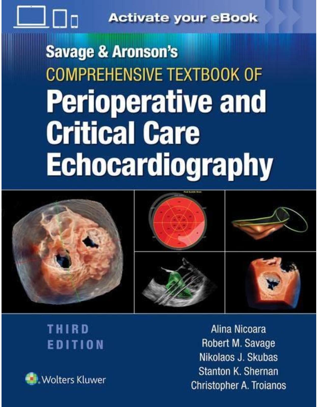
Comprehensive Textbook of Perioperative and Critical Care Echocardiography
Livrare gratis la comenzi peste 500 RON. Pentru celelalte comenzi livrarea este 20 RON.
Description:
Thoroughly revised to reflect new advances in the field, Savage & Aronson’s Comprehensive Textbook of Perioperative and Critical Care Echocardiography, Third Edition, remains the definitive text and reference on transesophageal echocardiography (TEE). Edited by Drs. Alina Nicoara, Robert M. Savage, Nikolaos J. Skubas, Stanton K. Shernan, and Christopher A. Troianos, this authoritative reference covers material relevant for daily clinical practice in operating rooms and procedural areas, preparation for certification examinations, use of echocardiography in the critical care setting, and advanced applications relevant to current certification and practice guidelines.
Table of Contents:
Section I: Fundamentals of Perioperative Echocardiography
Chapter 1. Physics of Ultrasound
Introduction
Echocardiography and the Properties of Ultrasound
Propagation of Ultrasound: Reflection, Refraction, and Attenuation
Specular and Nonspecular Reflection
Acoustic Impedance
Attenuation
Ultrasound Interactions and the Challenges to Signal Processing
The Piezoelectric Effect and Ultrasound
Ultrasound Transducers: The Basics
How Is the Generated Ultrasound Beam Steered?
Ultrasound Beam Geometry
Pulse-Echo Operation: Time Equals Distance
Image Display Formats
2D Echocardiography: Frames and Frame Rate
Tissue Harmonic Imaging
Contrast Echocardiography
Doppler Echocardiography
Blood Flow Hemodynamics, Flow Velocity Profiles, and Doppler Echocardiography
Doppler Principle and Doppler Frequency Shift
Continuous Wave Doppler Echocardiography
Pulsed Wave Doppler Echocardiography
High PRF Mode and Multigate Doppler
Graphical Display of Doppler Frequency Spectra
Tissue Doppler Echocardiography
Color Flow Doppler Echocardiography
Instrumentation Factors in Color Flow Doppler Imaging
Conclusions
Chapter 1: Multiple Choice Questions
Chapter 2. Doppler Echocardiography
Introduction
Blood flow Hemodynamics, flow Velocity Profiles, and Doppler Echocardiography
The Doppler Principle and Doppler Frequency Shift
Continuous Wave Doppler Echocardiography
Pulsed Doppler Echocardiography
High PRF Mode and Multigate Doppler
Graphical Display of Doppler Frequency Spectra
Tissue Doppler Echocardiography
Color Flow Doppler Echocardiography
Instrumentation Factors in Color Flow Doppler Imaging
CW, PW, and Color Flow Doppler Compared
Conclusions
Chapter 2: Multiple Choice Questions
Chapter 3. Imaging Artifacts and Diagnostic Pitfalls
Artifacts
Missing Structures
Degraded Images
Falsely Perceived Objects
Misregistered Locations
Artifacts in Doppler Analysis
Artifacts in Three-Dimensional Ultrasound
Stitching (Reconstruction) Artifacts
Dropout Artifacts
Blurring (Amplification) Artifacts
Blooming Artifact
Railroad Artifacts
Pitfalls
Right Atrium
Right Ventricle
Left Atrium
Left Atrioventricular Groove
Left Ventricle
Aortic Valve
Aorta
Pericardium
Conclusion
Chapter 3: Multiple Choice Questions
Chapter 4. Optimizing Two-Dimensional and Three-Dimensional Echocardiographic Images
Introduction
The Impact of Ultrasound Instrumentation on Image Generation and Display
Master Synchronizer (Clock)
Transducers and Spatial Resolution
Temporal Resolution and Real-Time Imaging
Amplification and Time Gain Compensation
Preprocessing
Analog-to-Digital Converters
Scan Conversion and Storage in Computer Memory
Postprocessing
Image Display, Recording, and Storage
Optimizing Three-Dimensional (3D) Images
Data Acquisition
Image Display
Specific Strategies to Optimize 3D Echo Imaging of the Cardiac Chambers
Specific Strategies to Optimize 3D Echo Imaging of the Valves
Conclusions
Chapter 4: Multiple Choice Questions
Chapter 5. Surgical Anatomy of the Heart
Fibrous Skeleton of the Heart
Cardiac Ventricles
Tricuspid Valve
Pulmonic Valve
Mitral Valve Apparatus
Mitral Valve Leaflets: Carpentier-SCA Terminology
Mitral Valve Apparatus—Duran Terminology
Aortic Root
Coronary Anatomy
Conclusion
Chapter 5: Multiple Choice Questions
Chapter 6. Comprehensive and Basic Perioperative TEE Examination
Prevention of TEE Complications
Intraoperative TEE Indications
Optimizing Image Quality
Comprehensive TEE Examination
General Considerations
TEE Probe Insertion
TEE Probe Manipulation
Left Atrium and Pulmonary Veins
Mitral Valve Midesophageal Views
Aortic Valve Midesophageal Views
Left Ventricle Midesophageal Views
Right Heart Midesophageal Views
Left Ventricle Transgastric Views
Mitral Valve Transgastric Views
Right Heart Transgastric Views
Aortic Valve Transgastric Views
TEE Examination of the Thoracic Aorta
Basic Perioperative TEE Examination
Limitations
Perioperative TEE Indications for Noncardiac Surgery
Training
Chapter 6: Multiple Choice Questions
Chapter 7. Indications for Intraoperative Transesophageal Echo
Introduction
Cardiac Surgery
Hemodynamic Instability
Trauma
Pulmonary Embolism
Venous Air Embolism
Lung Transplantation
Liver Transplantation
Renal Cell Carcinoma With IVC Extension
Adults With Congenital Heart Disease Undergoing Noncardiac Surgery
Complications
Chapter 7: Multiple Choice Questions
Chapter 8. Organization of a Perioperative Echocardiographic Service: Personnel, Equipment, Maintenance, Safety, Infection, Economics, and Continuous Quality Improvement
Personnel
Equipment and Maintenance
Safety
Complications Related to the Gastrointestinal System
Complications Related to Compression of Adjacent Structures
Contraindications
Cleaning and Disinfection of TEE Probes
Economics of an Intraoperative Echo Service
TEE Reporting and Billing
Billing for TEE
Billing Codes for Intraoperative TEE
Use of Modifiers
Bundling Issues
Diagnosis Codes
Credentialing
Assessing Quality in a Perioperative TEE Service
Chapter 8: Multiple Choice Questions
Chapter 9. Education and Training in Perioperative Echocardiography
Perioperative Echocardiography Training Programs
Resources for Perioperative Echocardiography Education
Certification in Perioperative Echocardiography
Chapter 9: Multiple Choice Questions
Chapter 10. Assessment of Global Left Ventricular Systolic Function
Normal Anatomy and Physiology of the Left Ventricle
Phases of Ventricular Systole
Left Ventricular Systolic Function
Two-Dimensional Examination of the Left Ventricle
Ejection Phase Indices of Left Ventricular Performance
Cardiac Output
Fractional Shortening
Fractional Area Change
Ejection Fraction
Calculating Preload and Afterload
Preload
Afterload
Systolic Index of Contractility (dP/dT)
Myocardial Performance Index
Tissue Doppler Imaging
Myocardial Strain by Tissue Doppler Imaging
Myocardial Deformation by Speckle Tracking
Three-Dimensional Evaluation of Global Ventricular Performance
Chapter 10: Multiple Choice Questions
Chapter 11. Regional Left Ventricular Systolic Function
Introduction
Segmental Model of the Left Ventricle
Distribution of Coronary Artery Anatomy
Clinical Application of Regional Wall Motion Analysis
Regional Ventricular Function and Ischemia Detection
Regional Ventricular Function and Nonischemic Conditions
Assessment of Myocardial Viability
Dobutamine Stress Echocardiography
Contrast-Enhanced Echocardiography
Other Echocardiographic Techniques for Assessing Regional Ventricular Function
Tissue Doppler Imaging
Speckle Tracking
Three-Dimensional Echocardiography
Chapter 11: Multiple Choice Questions
Chapter 12. Assessment of Right Ventricular Function
Introduction
Structure and Function of the Right Ventricle
Transesophageal Echocardiographic Scan Planes for Assessing the Right Ventricle
Right Ventricular Hypertrophy and Dilatation
Right Ventricular Pressure and Volume Overload and the Ventricular Septum
Right Ventricular Systolic Function—Qualitative and Regional Assessment
Right Ventricular Global Systolic Function—Quantitative Echocardiographic Assessment
Longitudinal Motion
Two-Dimensional and Three-Dimensional RV Quantification
Dimensionless RV Quantification
Right Ventricular Function—Hemodynamic Assessment
Right Ventricular Diastolic Function
Diastolic Function: Right Atrial Dimensions
Chapter 12: Multiple Choice Questions
Chapter 13. Assessment of Diastolic Function in the Perioperative Setting
Introduction
Clinical Importance of Diastolic Dysfunction
Physiology of Diastole and Pathophysiology of Dysfunction
LV Relaxation
LV Compliance
Left Atrial Function
Mitral Annular Excursion
Echocardiographic Evaluation of Left Ventricular Diastolic Function
Transmitral Inflow Velocities
Stage I: Impaired Relaxation Pattern
Stage II: Pseudonormal Pattern
Stage III: Restrictive Pattern
Pulmonary Venous Flow
Mitral Annulus Tissue Doppler Imaging
Color M-Mode Doppler: Propagation Velocity
Left Atrial Strain
Grading of Diastolic Dysfunction
Limitations of Current Techniques of Echocardiographic Assessment of Diastolic Dysfunction
Emerging Techniques for Assessing LV Diastolic Function
Right Ventricular Diastolic Function
Clinical Implications of Diastolic Dysfunction in the Perioperative Setting
Conclusions
Chapter 13: Multiple Choice Questions
Chapter 14. Assessment of the Mitral Valve
Anatomy of the Mitral Valve
Structural Integrity of the Mitral Valve
Mitral Regurgitation
Ischemic Mitral Regurgitation
Severity Estimation
Two-Dimensional Echocardiography
Qualitative Techniques
Continuous Wave Doppler Signal
Peak E-Wave Velocity
Semiquantitative Techniques
Spatial Area Mapping—Jet Area
Pulmonary Venous Waveform Patterns
Quantitative Techniques
Vena Contracta
Effective Regurgitant Orifice Area
Regurgitant Volume and Regurgitant Fraction
Three-Dimensional Echocardiography
Mitral Stenosis
Two-Dimensional Echocardiography
Mitral Valve Scoring Systems
Severity Estimation
Valve Area Calculations
Nonrheumatic Etiologies of Mitral Stenosis
Cor Triatriatum
Chapter 14: Multiple Choice Questions
Chapter 15. Assessment of the Aortic Valve
Introduction
Anatomy of the Aortic Valve
Assessing the Aortic Valve With Transesophageal Echocardiography
Midesophageal AV Short-Axis (ME AV SAX) View
Midesophageal AV Long-Axis (ME AV LAX) View
Deep Transgastric Five Chamber View
Transgastric Long-Axis View
Three-Dimensional Imaging of the AV
Aortic Stenosis
Pathophysiology
Quantification of Aortic Stenosis
Peak Aortic Valve Jet Velocity
Aortic Valve Mean Gradient Measurement
Aortic Valve Area
Indexed Aortic Valve Area
Velocity Ratio and Dimensionless Index (VTI Ratio)
Limitations With the Velocity Ratio and Dimensionless Index
Low-Flow, Low-Gradient Aortic Stenosis
Low-Flow, Low-Gradient Aortic Stenosis With Decreased LV Ejection Fraction
Low-Flow, Low-Gradient Aortic Stenosis With Preserved LV Ejection Fraction
Aortic Regurgitation
Pathophysiology
Quantification of Aortic Regurgitation
Chapter 15: Multiple Choice Questions
Chapter 16. Assessment of the Tricuspid and Pulmonic Valves
Introduction
Structure and Function of the Tricuspid and Pulmonic Valves
Structure and Function of the Tricuspid Valve
Structure and Function of the Pulmonic Valve
Transesophageal Echocardiographic Evaluation of the Tricuspid and Pulmonic Valves
Two-Dimensional and Color Flow Doppler Examination
Spectral Doppler Examination
Three-Dimensional Examination
Tricuspid Regurgitation
Pulmonic Regurgitation
Tricuspid Stenosis
Pulmonic Stenosis
Congenital Diseases of the Tricuspid and Pulmonic Valves
Ebstein Anomaly
Tricuspid Atresia
Congenital Anomalies of the Pulmonic Valve
Acquired Diseases of the Tricuspid and Pulmonic Valves
Functional Tricuspid Regurgitation
Endocarditis
Carcinoid Heart Disease
Rheumatic Heart Disease
Other Pathologic Conditions
The Ross Procedure
Chapter 16: Multiple Choice Questions
Chapter 17. Prosthetic Heart Valves
Types of Prosthetic Valves
Mechanical Valves
High-Profile Valves
Low-Profile Valves
Medtronic Hall Valve
OmniScience Valve
Gott-Daggett Valve
St. Jude Valve
Carbomedics Valve
On-X Valve
Bioprosthetic Valves
Mitral Homograft Valves
Aortic Homograft Valves
Xenografts
Hancock Porcine Valve
Carpentier-Edwards Porcine Valves
Ionescu-Shiley Valve
Carpentier-Edwards Perimount
Carpentier-Edwards Perimount Magna, Magna Ease, and Inspiris
St. Jude Medical Biocor and Trifecta
Sorin Mitroflow
Medtronic Mosaic and Mosaic Ultra
Echocardiographic Evaluation of Prosthetic Heart Valves
Standard Echo Exam
Pressure Gradients
Effective Orifice Area
Pressure Half-Time
Normal Prosthetic Valve Flow Patterns
Mechanical Valves
Physiologic Regurgitation
Pressure Recovery
Imaging Artifact With Prosthetic Valves
Star-Edwards Valve (Mitral Position)
Disc Valves
Bjork-Shiley Valve (Mitral Position)
Medtronic-Hall Valve (Mitral Position)
Bileaflet Valves
Stented Bioprosthetic Valves
Stentless Bioprosthetic Valves
Aortic Valve Homografts
Three-Dimensional TEE
Echocardiographically Detectable Pathology
Structural vs Nonstructural Damage
Prosthetic Valve Stenosis
Patient Prosthesis Mismatch
Prosthetic Valve Regurgitation
Prosthetic Valve Endocarditis
Associated Systemic Complications
Thromboembolism
Hemorrhage
Hemolysis
Chapter 17: Multiple Choice Questions
Chapter 18. Assessment of Cardiac Masses
Approach/Structures
Inferior Vena Cava/Superior Vena Cava/Right Atrium
Right Ventricle/Pulmonary Artery
Pulmonary Veins/Left Atrium/Left Atrial Appendage
Left Ventricle
Aorta
Valves
Pathology
Inferior Vena Cava/Superior Vena Cava/Right Atrium
Right Ventricle/Pulmonary Artery
Pulmonary Veins/Left Atrium/Left Atrial Appendage
Left Ventricle
Aorta/Valves
Chapter 18: Multiple Choice Questions
Chapter 19. Pericardial Diseases
Pericardial Anatomy
Pericardial Physiology
Macrophysiology
Microphysiology
Respirophasic Variation
Pericardial Pathology
Congenital Pericardial Defects
Pericarditis
Pericardial Effusion and Tamponade
Constrictive Pericarditis
Pericardial Masses
Chapter 19: Multiple Choice Questions
Section II: Advanced Perioperative Applications of Echocardiography in Cardiothoracic Surgery
Chapter 20. Cannulation and Perfusion Strategies for Cardiopulmonary Bypass
Peripheral Venous Cannulation
Femoral Arterial Cannulation
Endovascular Aortic Cross-Clamp
Percutaneous Coronary Sinus Catheters and Pulmonary Artery Vents
Percutaneous Coronary Sinus Catheters
Imaging the Coronary Sinus
Placement
Confirmation
Percutaneous PA Vents
Chapter 20: Multiple Choice Questions
Chapter 21. Epiaortic and Epicardial Imaging
Epicardial Imaging
Indications
Imaging Guidelines
Epiaortic Imaging
Indications
Imaging Guidelines
Grading Systems
Implications for Surgical Management
Imaging Technique
Epicardial Views
Epiaortic Views
Chapter 21: Multiple Choice Questions
Chapter 22. Assessment of Perioperative Hemodynamics
Doppler Measurements of Stroke Volume and Cardiac Output
Calculation of Stroke Volume
Calculation of Cardiac Output
Calculation of LVOT Stroke Volume (Fig. 22.7)
Calculation of Transaortic Valve Stroke Volume (Fig. 22.8)
Calculation of Pulmonary Annulus Stroke Volume (Fig. 22.9)
Calculation of RVOT Stroke Volume (Fig. 22.10)
Calculation of Transmitral Stroke Volume (Fig. 22.11)
Doppler Measurement of Pulmonary-To-Systemic Flow Ratio (Qp/Qs) (Fig. 22.12)
Doppler Assessment of Regurgitation
Regurgitant volume and fraction—Volumetric Method (Fig. 22.13)
Assessment of Mitral Regurgitation (Fig. 22.14)
Assessment of Aortic Regurgitation (Fig. 22.15)
Proximal Convergence Method
Simplified Proximal Convergence Method
Doppler Measurement of Pressure Gradients
Doppler Determination of Valve Area
Continuity Equation (Fig. 22.22)
Continuity Equation in Aortic Stenosis (Fig. 22.23)
Continuity Equation in Mitral Regurgitation (the Flow Convergence Method)
Continuity Equation in Mitral Stenosis (the Flow Convergence Method)
Pressure Half-time
Doppler Determination of Intracardiac Pressures
Estimation of RVSP (Fig. 22.29)
Estimation of PASP
Estimation of PADP (Fig. 22.30)
Estimation of MPAP
Estimation of LAP (Fig. 22.31)
Estimation of LVEDP (Fig. 22.32)
Doppler Measurement of DP/DT
Doppler Measurement of Vascular Resistance
Chapter 22: Multiple Choice Questions
Chapter 23. Assessment and Surgical Considerations in Ischemic/Functional Mitral Valve Surgery
Introduction
Pathophysiology
Acute Ischemic Mitral Regurgitation
Chronic Ischemic Mitral Regurgitation
Assessment of Ischemic/Functional Regurgitation Severity
Hemodynamic Considerations in Assessing MR Severity
Surgical Management
Predictors of Failed MV Repair in Ischemic MR
Dyssynchrony and Chronic Ischemic MR
Percutaneous Interventions for Ischemic/Functional MR
Chapter 23: Multiple Choice Questions
Chapter 24. Surgical Considerations and Assessment in Nonischemic Mitral Valve Surgery
Historical Perspectives
Importance of Intraoperative Echocardiography (IOE) in MV Surgery
Importance of Intraoperative EchocardiograpHy (IOE) MV Assessment in the Future of Cardiothoracic Anesthesia
Intraoperative Echocardiography and Critical Issues in MV Surgery
Organization of Chapter
Mitral Valve Apparatus
Normal Anatomy of the Mitral Valve Apparatus
Fibrous Skeleton
Mitral Annulus
Mitral Valve Leaflets
Chordae Tendinae
Papillary Muscles
Left Ventricle
Nomenclature of the Mitral Valve Apparatus
Correlation With Imaging Planes: the 2D Examination of the Mitral Valve
Midesophageal Four-Chamber MV and Variations
Midesophageal Four-Chamber MV (ME 4Ch MV)
Midesophageal Five-Chamber MV (ME 5Ch MV)
Lower-Esophageal Four-Chamber MV (LE 4Ch MV)
Midesophageal Commissural MV and Variations
Midesophageal Commissural MV (ME Com MV)
Midesophageal Commissural Right MV (ME ComR MV)
Midesophageal Commissural Left MV (ME ComL MV)
Midesophageal Two-Chamber MV and Variations
Midesophageal Two-Chamber MV (ME 2Ch MV)
Midesophageal Two-Chamber Right MV (ME 2ChR MV)
Midesophageal Two-Chamber Left MV (ME 2ChL MV)
Midesophageal Long-Axis MV and Variations
Midesophageal Long-Axis MV (ME LAX MV)
Midesophageal Long-Axis Right MV (ME LAXR MV)
Midesophageal Long-Axis Left MV (ME LAXL MV)
Transgastric Basal Short Axis (TG SAXB)
Transgastric Two Chamber (TG 2Ch)
Intraoperative Examination Approach
Principles of Intraoperative Examination (Table 24.5)
Intraoperative Echocardiography Examination and Outcomes
Pre–Cardiopulmonary Bypass (Pre-CPB IOE)
The Pre-CPB IOE Exam and Outcome Studies (Table 24.7)
New Clinical Information
Determine Mechanism and Probability of Repair
Variances With Preoperative MV Dysfunction Severity
Predicting Complications
Systolic Anterior Motion With LVOT Obstruction (LVOTO)
Periannular Annular Disruption
LV Dysfunction
Cannulation-Perfusion and Myocardial Protection Strategy
Post-CPB Exam and Outcomes
Post-CPB Clinical Issues (Tables 24.8, 24.9, 24.17, and 24.18)
Post-CPB Complications
Post-CPB Outcomes
Mitral Stenosis
Micro-Air and LV Function
Aortic Dissection
Post-CPB LV Dysfunction
Annular Disruption Following Surgical Debridement
Iatrogenic Shunts and Fistulas
Perivalvular MR Following MVR
Systematic IOE Examination in MV Surgery
Abbreviated Overview Exam (Table 24.10; Fig. 24.24) (II)
Focused Diagnostic IOE Exam in MV Surgery (Table 24.11)
General Comprehensive Exam
Pathology
Mitral Regurgitation
Etiology and Mechanisms of Mitral Regurgitation (Tables 24.12-24.14)
Mechanisms of Mitral Regurgitation (Fig. 24.63; Tables 24.13-24.15)
Type I: Normal Leaflet Motion (Fig. 24.63)
Type II: Excessive Leaflet Motion (Fig. 24.63)
Type III: Restricted Leaflet Motion (Fig. 24.63)
Intraoperative Echocardiography and Mechanisms of MVAp Dysfunction
Type I: Normal Leaflet Motion (Fig. 24.63)
Type II: Excessive Leaflet Motion (Fig. 24.63)
Type III Restricted Leaflet Motion (Fig. 24.63)
Comparison of IOE Exam and Direct Surgical Inspection (Tables 24.14 and 24.15)
Surgical Technique and IOE Characterization (Tables 24.14 and 24.15)
IOE Assessment of MR Severity (Tables 24.16-24.18)
Two-Dimensional IOE
Doppler Assessment of MR
Color-Flow Doppler Maximal Jet Area (MJA)
Vena Contracta Diameter (VCD)
Proximal Flow Convergence
Simplified PISA Method (Fig. 24.65; Table 24.18)
Continuous Wave Doppler
Pulse Wave Doppler
Mitral Stenosis
Severity Assessment (Table 24.21)
IOE Two-Dimensional Assessment of Severity of MS
Doppler Methods of Severity Assessment
IOE Findings Associated With MV Dysfunction
Associated Valvular or Coexisting Cardiac Pathology
Aortic Stenosis or Regurgitation in Patient for MV Surgery
Endocarditis of the Mitral and Aortic Valves
Mitral Valve Procedures
Introduction
Mitral Valve Repair Historical Perspective
MV Repair for Rheumatic Disease
MV Repair for Endocarditis
MV Repair for Ischemic Heart Disease
MV Repair for Myxomatous MV Disease
Clinical Guidelines for MV Repair
Moderate MR in Patients for Aortic Valve Surgery
Mitral Valve Replacement
Transcatheter Approaches to Mitral Valve Disease
Pre-CPB IOE Exam
Post-CPB IOE Exam
Intraoperative Three-Dimensional Echocardiography of the Mitral Valve
Chapter 24: Multiple Choice Questions
Chapter 25. Assessment and Surgical Considerations in Aortic Valve Surgery
Role of TEE During Aortic Valve Surgery
Clinical Dilemmas During Aortic Valve Surgery
Moderate Aortic Stenosis
Low-Gradient Aortic Stenosis
Hemodynamic Management With Altered Ventricular Compliance
Focused Intraoperative Echocardiography Examination for Aortic Valve Surgery
Specific Aortic Valve Procedures
Aortic Valve Replacement
Stentless and Sutureless Prosthetic Aortic Valves
Aortic Valve Repair
Combined Aortic Valve and Aortic Root Surgery
Ross Procedure
Commando Procedure
Cor-Knot
Chapter 25: Multiple Choice Questions
Chapter 26. Assessment and Surgical Considerations During Open and Endovascular Thoracic Aortic Surgery
Anatomy of the Thoracic Aorta
Transesophageal Echocardiography in the Examination of the Aorta
Introduction
Transesophageal Echocardiography Views
Overview of Thoracic Aortic Diseases
Aortic Aneurysms
Aortic Dissections
Traumatic Injuries
Intramural Hematoma
Atherosclerotic Disease
Coarctation and Other Congenital Anomalies
Intraoperative Role of TEE in Aortic Disease
Thoracic Aortic Aneurysm
Thoracic Aortic Atheroma
Type A Aortic Dissections
Confirmation of the Diagnosis
Assessment of the Aortic Root, Aortic Valve, and Coronary Ostia
Assessment of Global and Regional Myocardial Function
Monitoring for LV Distention Cooling
Postrepair Assessment
Type B Aortic Dissections Open Surgical Repairs
Thoracic Endovascular Aortic Repair Procedure
Guiding Stent-Graft Placement
Endoleaks Classification
Detecting Endoleaks
Monitoring Cardiac Performance During TEVAR
Limitations of TEE
Role of Intravascular Ultrasound
Summary and Future Directions
Chapter 26: Multiple Choice Questions
Chapter 27. The Assessment of a Patient With Endocarditis
Epidemiology and Risk Factors
Diagnosis
Echocardiography
Vegetation
Complications Secondary to IE
Valvular Regurgitation
Abscess and Perivalvular Involvement
Pseudoaneurysms and Intracardiac Fistulae
The Prognostic Utility of Echocardiography and IE
The Role of 3D Echocardiography
Surgical Indications for Infective Endocarditis
Intraoperative Assessment
Conclusion
Chapter 27: Multiple Choice Questions
Chapter 28. Hemodynamic Assessment While Weaning Off Cardiopulmonary Bypass
Introduction
Epidemiology
A Proposed Focused TEE Examination During Weaning Off CPB
Echocardiographic Associations for Hemodynamic Aims
Preload
Regional LV function
Difficult Weaning Off CPB
Preload
LV Failure
RV Failure
Structural Defects
Inappropriate Vasodilation
Conclusion
Chapter 28: Multiple Choice Questions
Section III: Advanced Perioperative Applications of Echocardiography in End-Stage Heart and Lung Failure
Chapter 29. Echocardiographic Assessment of Cardiomyopathies
Dilated Cardiomyopathy
Echocardiographic Features
Hypertrophic Cardiomyopathy
Echocardiographic Features
Restrictive Cardiomyopathy
Echocardiographic Features
Arrhythmogenic Right Ventricular Cardiomyopathy
Echocardiographic Features
Unclassified Cardiomyopathies
Role of Echocardiography: Prognostication and Treatment
Summary
Chapter 29: Multiple Choice Questions
Chapter 30. Assessment and Surgical Consideration in Pericardiectomy
The Normal Pericardium
Clinical Perspectives
Pericardial Clinical Syndromes
Epidemiology and Etiologies of Constrictive Pericarditis
Clinical Presentation of Constrictive Pericarditis
Constrictive Pericarditis—Echocardiographic Assessment
Two-Dimensional Imaging and M-Mode
Doppler Echocardiography and Strain Assessment
Diagnostic Criteria
Other Multimodality Imaging
Pericardiectomy—Surgical Considerations
Indications
Surgical Approach
Intraoperative Echocardiography
Outcomes
Conclusion
Chapter 30: Multiple Choice Questions
Chapter 31. Assessment and Surgical Considerations in Cardiac Transplantation
Transesophageal Echocardiography in Cardiac Transplantation
The Role of TEE in Cardiac Donor Screening
Intraoperative Monitoring in the Pretransplantation Period
Left Ventricular Volume
Left Ventricular Contractility
Intracavitary Thrombus
Atherosclerosis of the Ascending Aorta and Aortic Arch
Right Ventricular Dilation or Hypertrophy
Assessment of Left Ventricular Assist Device Explant
Hemodynamic Calculations
Intraoperative Monitoring in the Posttransplantation Period
Assisting With Venting and De-airing Maneuvers
Separation From Cardiopulmonary Bypass
Assessment of the Left Ventricle Systolic Function
Assessment of LV Diastolic Function
Assessment of the Right Ventricle
Etiology, Pathophysiology, and Diagnosis of Acute RV Dysfunction
Management of Acute RV Dysfunction
Assessment of the Atria and Atrial Anastomoses
Assessment of the Vena Cava Anastomoses
Assessment of the Pulmonary Artery Anastomosis
Assessment of the Pulmonary Venous Anastomoses
Assessment of the Atrioventricular Valves
Management of Early Postoperative Hemodynamic Abnormalities in the Intensive Care Unit
Postoperative Follow-up Studies of Cardiac Allograft Function
New Advances in Heart Transplantation
Chapter 31: Multiple Choice Questions
Chapter 32. Assessment and Surgical Considerations in Lung and Heart-Lung Transplantation
Lung Transplantation
Surgical Considerations
TEE Assessment of Patients Undergoing Lung Transplantation
Right Ventricular Function
Intracardiac Shunts
Vascular Anastomoses
Posttransplantation
TEE Guidance for Mechanical Circulatory Support
Heart-Lung Transplantation
Summary
Chapter 32: Multiple Choice Questions
Section IV: Advanced Perioperative Applications of Echocardiography in Structural Heart Disease
Chapter 33. Assessment and Procedural Considerations During Transcatheter Aortic Valve Replacement
Related Anatomy
Prosthesis
Periprocedural Imaging for Tavr
Preprocedural Imaging
Deployment
Complications
Chapter 33: Multiple Choice Questions
Chapter 34. Assessment and Considerations During Left Atrial Appendage Closure Procedures
Left Atrial Appendage Anatomy
The Watchman LAA Occluder
Preprocedural Imaging
Procedural Guidance
Baseline Comprehensive Assessment
Left Atrial Appendage Sizing
Transseptal Puncture
Accessing the LAA
Device Delivery
Post Deployment Assessment
Device Release and Assessment
Complications
Postprocedural Management and Follow-up
Future Directions
Chapter 34: Multiple Choice Questions
Chapter 35. Assessment and Considerations During Transcatheter Mitral Valve Procedures
Mitral Valve Pathology
Anatomical Implications for Transcatheter Mitral Valve Therapy
Decision-making in Mitral Regurgitation: Surgical vs Transcatheter Repair or Replacement
Role of Echocardiography in Transcatheter Mitral Interventions
Transseptal Puncture
Transcatheter Mitral Valve Repair Techniques
Transcatheter Edge-to-Edge Repair
Mitraclip
PASCAL Leaflet Plication Device
Percutaneous Annuloplasty Techniques
Percutaneous Chordal Repair Techniques
Transcatheter Mitral Valve Replacement
General Considerations and Echocardiographic Principles
Potential Complications
Transcatheter Mitral Valve Paravalvular Leak Closure
Transcatheter Therapy for Mitral Stenosis
Percutaneous Balloon Valvuloplasty
Considerations Surrounding the Use of Three-Dimensional Echocardiography and Other Imaging Adjuncts
Acknowledgment
Chapter 35: Multiple Choice Questions
Chapter 36. Assessment and Considerations During Transcatheter Tricuspid Valve Repair and Replacement
Lessons Learned from Surgery: Avoiding the Mistakes of the Past
Current Recommendations
Outcomes of Surgical Intervention for TR
Efficacy and Durability of Tricuspid Valve Repair Techniques
Tricuspid Valve Replacement
Pre-procedural Imaging for Transcatheter Tricuspid Valve Interventions: Cardiac Magnetic Resonance, Computed Tomography and Echocardiography
Cardiac Magnetic Resonance
Right Ventricular Size and Function
Tissue Characterization and Fibrosis Imaging
Tricuspid Valve Assessment
Cardiac Computed Tomography Angiography
Evaluation of Right Heart and Tricuspid Valve Anatomy
Pretranscatheter Tricuspid Valve Intervention Planning
Transthoracic Echocardiography
TR Severity Assessment
Annular Dilation
Papillary Muscle Displacement and Leaflet Tethering
Right Ventricular Size and Function
RV-RA Gradient and Pulmonary Hypertension
Transesophageal Echocardiography
Three-Dimensional (3D) Echocardiography
Transcatheter Tricuspid Valve Devices
Annular Devices
Leaflet Coaptation Devices
Heterotopic Transcatheter Tricuspid Valve Replacement
Orthotopic Transcatheter Tricuspid Valve Replacement
Chapter 36: Multiple Choice Questions
Chapter 37. Assessment and Considerations for Atrial Septal Defect Closure
Anatomy of the Interatrial Septum
Echocardiographic Anatomy of the Interatrial Septum
ASD Subtype Amenable to Percutaneous Closure
Secundum Atrial Septal Defect Closure
Indications
Suitability for Percutaneous Closure
ASD Size, Shape, and Number of Defects
Length of Rims
Patent Foramen Ovale Closure
Indications
Anatomic Considerations
Choice of Device
Step by Step Guidance for Secundum ASD Percutaneous Closure
STEP 1: Rule Out Contraindications to the Procedure
STEP 2: Confirm ASD Anatomy and Suitability for Device Closure
STEP 3: Device Positioning
STEP 4: Rule Out Immediate Complications
Guidance for PFO Closure
Complications
Device Erosion
Embolization
Wire Fracture
Procedural Guidance Using Intracardiac Echocardiography
Chapter 37: Multiple Choice Questions
Section V: Advanced Perioperative Applications of Echocardiography During Electrophysiologic Procedures
Chapter 38. Procedural Intracardiac Ultrasound Imaging in Ablation of Arrhythmias
Imaging Technology
Radial Intracardiac Echocardiography Technology
Phased Array Technology
Insertion Technique
Basic Imaging
Home View
Cavotricuspid Isthmus View
Coronary Sinus View
Right Ventricular Outflow Tract
Septal Views
Left Pulmonary Veins
Right Pulmonary Veins
LV Structures
Pulmonary Artery Views
Fluoroless Transseptal Puncture
Technique
Atrial Fibrillation Ablation
Atrial Flutter Ablation
Ventricular Tachycardia Ablation
Ice-Guided Watchman Implant
Complication Prevention
Pericardial Effusion
Radiofrequency Ablation
Tissue Heating
Ice Guidance for Other Procedures
Coronary Sinus Catheter Placement
Interventricular Septal Puncture
Chapter 38: Multiple Choice Questions
Chapter 39. Insertion of Rhythm Management Devices and Cardiac Resynchronization Therapy
Introduction
Rationale for CRT
Indications for CRT
Echocardiography for Selection of CRT Responders
Cardiac Mechanical Dyssynchrony
Mechanical Discoordination
Myocardial Viability and CRT Response
Contribution of RV Function to CRT Response
Specific Echocardiographic Evaluation of Dyssynchrony
Speckle Tracking Echocardiography for Strain
Novel TDI-Based Methods
Three-Dimensional Echocardiography
Additional Echocardiographic Assessment Prior to CRT
Intraprocedural Echocardiographic Guidance
Complications of CRT Implantation
Chapter 39: Multiple Choice Questions
Chapter 40. Transvenous Lead Extraction
Introduction
Pathology
Traumatic Tricuspid Valve Injury
Infective Endocarditis
Cardiac Perforation and Tamponade
Embolic Events
Chapter 40: Multiple Choice Questions
Section VI: Diagnostic Echocardiography and Ultrasound in Critical Care Settings
Chapter 41. Overview of Transesophageal Echocardiography and Transthoracic Echocardiography in Critical Care
Introduction
Terminology and Use
Rationale and Approach to CCE
Cardiac Ultrasound Protocols
Ultrasound Equipment
Chest Wall Windows
Essential Transthoracic Views
The Parasternal Window
Parasternal Short Axis
Apical Four-Chamber View (A4C) and Five-Chamber View (A5C)
Apical Tow-Chamber (A2C) and Apical Three-Chamber View (A3C)
The Subcostal Window
Conclusion
Chapter 41: Multiple Choice Questions
Chapter 42. Hemodynamic Assessment of the Critically Ill Patient
Introduction
Left Ventricular Function
Right Ventricular Function
Stroke Volume and Cardiac Output
Left Ventricular Outflow (LVOT) Obstruction
Cardiac Arrest
Intracardiac Filling Pressures and Volume Status
Left Heart Filling Pressures
Right Heart Filling Pressures
Overall Intravascular Volume Status
Valvular Function and Disease
Valvular Stenosis
Aortic Valve Stenosis
Mitral Valve Stenosis
Valvular Insufficiency
Aortic Valve Regurgitation
Mitral Valve Regurgitation
Prosthetic Valve Dysfunction
Pulmonary Artery Pressures
Pulmonary Embolism
Pericardial Disease
Intracardiac and Intrapulmonary Shunts
Acute Coronary Syndrome and Its Complications
Complications After Cardiac Surgery
Mechanical Circulatory Support Devices
Intraaortic Balloon Pump
Impella
Venoarterial Extracorporeal Membrane Oxygenation
Left Ventricular Assist Devices
Conclusion
Chapter 42: Multiple Choice Questions
Chapter 43. Rescue Echocardiography in the Unstable Patient
Left Ventricular Failure
Right Ventricular Failure
Severe Valve Disease
Systolic Anterior Motion
Pericardial Tamponade
Hypovolemia
Low Arterial Tone and Hemodynamics
Chapter 43: Multiple Choice Questions
Chapter 44. Acute and Chronic Pulmonary Hypertension
Introduction
Definition of Pulmonary Hypertension
Classification of Pulmonary Hypertension Using Echocardiography
Tricuspid Regurgitation
Pulmonary Regurgitation
Acceleration Time
Pulmonary Vascular Resistance
Right Ventriculo-Pulmonary Arterial Coupling
The Anatomic and Functional Correlates of Pulmonary Hypertension
Acute and Chronic Pulmonary Embolic Disease
Chapter 44: Multiple Choice Questions
Chapter 45. Assessment of the Lung and Thoracic Cavity in ICU
Introduction
Approach: the “P3-D Method”
Pleural Interface Integrity
Lung Parenchyma
Pleural Effusion and Diaphragm Motion
Normal Anatomic Findings
Ultrasonographic Pathological Findings
Pneumothorax
Probe Selection and Placement
Evaluation Process
Pulmonary Edema
Probe Selection and Placement
Evaluation
Acute Respiratory Distress Syndrome
Evaluation and Ultrasound Findings
Clinical Utility
Pneumonia
Evaluation and Ultrasound Findings
Limitations
Chapter 45: Multiple Choice Questions
Chapter 46. Assessment of Acute Kidney Injury and Renal Function
Introduction
Approach and Structures
Pathology
Pathophysiology of Acute Kidney Injury
Diagnosis and Evaluation of Acute Kidney Injury
Diagnosis and Prediction of Acute Kidney Injury Using Ultrasound
Renal Resistive Index
Renal Venous Assessment
Hepatic Vein and Portal Vein Assessment
Delineation of Acute Kidney Injury Etiology
Obstruction (Bladder, Hydronephrosis)
Volume Status and Renal Perfusion
Summary
Chapter 46: Multiple Choice Questions
Chapter 47. Vascular Access
Indications for Vascular Access
Central Venous Access
Arterial Access
Peripheral Access
Technical Considerations
Two-Dimensional vs Doppler Ultrasound
Transducer Probes
Style of Probe Use: “Free-Hand” vs Needle Guide
Short Axis vs Long Axis vs Oblique Axis
Ultrasound-Guided Central Venous Access
Evidence-Based Analysis for Ultrasound Use
Meta-analyses
Landmark- vs Ultrasound-Guided Technique
Anatomic Considerations
Variable Location of IJV
Overlap of IJV and CA
Other Anatomic Variables
Complications
Cannulation Site
Operator Experience
Patient Variables
Ultrasound Use in Pediatric Patients
Practice Guidelines for Ultrasound-Guided Central Venous Access
Ultrasound-Guided Arterial Access
Transesophageal Echocardiography for Vascular Access
Central Venous Access
Pulmonary Artery Catheters
Coronary Sinus Catheters
Acknowledgments
Chapter 47: Multiple Choice Questions
Chapter 48. Procedural Access in the Intensive Care Unit
Introduction
Structures
The Thoracic Cavity (Illustration of Normal Anatomy?)
The Pericardium and Pericardial Space
The Peritoneal Space
Pathology
Pleural Effusion
Pericardial Effusion
Ascites
Approach
Thoracentesis
Pericardiocentesis
Paracentesis
Chapter 48: Multiple Choice Questions
Section VII: Echocardiography in Noncardiac Settings
Chapter 49. Transesophageal Echocardiography for Nonthoracic Transplants
Introduction
Utility of TEE for Liver Transplantation
Patterns of TEE Utilization for Liver Transplantation and Barriers to Use
Phases of Liver Transplant/Postreperfusion Syndrome
Cardiovascular Comorbidities
TEE Findings During Liver Transplantation
Contraindications and Complications
Other Solid Organ Transplantation
Chapter 49: Multiple Choice Questions
Chapter 50. Intraoperative Imaging of the Inferior Vena Cava and Hepatic Vein by Transesophageal Echocardiography
Introduction
Structures: Anatomy and Physiology
Approach: TEE Imaging of the IVC and Hepatic Vein
2D Imaging Windows
IVC Variants
Doppler Interrogation of the Hepatic Veins
A-Wave
S-Wave
V-Wave
D-Wave
Pathology: Clinical Imaging of the IVC and Hepatic Veins
Renal Cell Carcinoma
Budd-Chiari Syndrome and Hepatic Vein Occlusion
Conclusion
Chapter 50: Multiple Choice Questions
Index
| An aparitie | 17 Oct. 2022 |
| Autor | Savage |
| Dimensiuni | 21.59 x 2.54 x 25.4 cm |
| Editura | LWW |
| Format | Hardcover |
| ISBN | 9781975102920 |
| Limba | Engleza |
| Nr pag | 816 |
| Versiune digitala | DA |
Clientii ebookshop.ro nu au adaugat inca opinii pentru acest produs. Fii primul care adauga o parere, folosind formularul de mai jos.